


生物技术通报 ›› 2023, Vol. 39 ›› Issue (5): 120-129.doi: 10.13560/j.cnki.biotech.bull.1985.2022-0999
钱榜1( ), 刘振东1, 赵印1, 李静1, PRAJAPATI Meera1,3, 李彦敏2, 孙跃峰1, 窦永喜1()
收稿日期:2022-08-05
出版日期:2023-05-26
发布日期:2023-06-08
通讯作者:
窦永喜,男,副研究员,研究方向:动物疫苗与分子免疫学;E-mail: douyongxi@caas.cn作者简介:钱榜,男,硕士研究生,研究方向:动物疫苗与分子免疫学;E-mail: QIANBang1993@outlook.com
基金资助:
QIAN Bang1(), LIU Zhen-dong1, ZHAO Yin1, LI Jing1, PRAJAPATI Meera1,3, LI Yan-min2, SUN Yue-feng1, DOU Yong-xi1()
Received:2022-08-05
Published:2023-05-26
Online:2023-06-08
摘要:
本研究旨在建立小反刍兽疫病毒H蛋白抗体的化学发光免疫分析检测方法。以H蛋白4个反应原性较好的B细胞表位串联后为检测抗原,在确定抗原包被量和血清稀释度,优化血清和酶标二抗反应时间的基础上,用ROC曲线分析确定检测临界值,建立了小反刍兽疫病毒H蛋白抗体化学发光免疫分析检测方法,然后对该方法进行敏感性、特异性和重复性评价。结果显示,建立的小反刍兽疫病毒H蛋白抗体化学发光免疫分析检测方法最佳抗原包被浓度为1.5×10⁻⁶ µg/孔,待检血清最佳稀释度为1∶100;血清及酶标二抗孵育时间均为10 min,检测方法的临界值为S/P=9.77%,批内及批间变异系数均小于10%;与口蹄疫、羊痘、蓝舌病及山羊传染性胸膜肺炎阳性血清不发生交叉反应;用建立的化学发光法和市售小反刍兽疫抗体cELISA试剂盒平行检测247份田间血清,两者相对符合率为94.33%,但化学发光法敏感性更高。研究结果表明,建立的小反刍兽疫病毒H蛋白抗体化学发光免疫分析方法灵敏、特异、稳定,可用于田间血清样品小反刍兽疫抗体检测。
钱榜, 刘振东, 赵印, 李静, PRAJAPATI Meera, 李彦敏, 孙跃峰, 窦永喜. 小反刍兽疫病毒H蛋白抗体化学发光免疫分析检测方法的建立[J]. 生物技术通报, 2023, 39(5): 120-129.
QIAN Bang, LIU Zhen-dong, ZHAO Yin, LI Jing, PRAJAPATI Meera, LI Yan-min, SUN Yue-feng, DOU Yong-xi. Establishment of Chemiluminescence Immunoassay for the Detection of Peste des Petits Ruminants Virus H Protein Antibodies[J]. Biotechnology Bulletin, 2023, 39(5): 120-129.
| 表位Epitope | 序列Sequence, N-C | 长度Length/AA |
|---|---|---|
| H123 | KFLNPDREYDFRDLR | 15 |
| H185 | GTGCLGRTVTRA | 8 |
| H362 | EANWVVPSTDVRDL | 14 |
| H487 | IRGPRGRCH | 9 |
表1 PPRV H蛋白4个反应原性最佳的B细胞表位序列
Table 1 4 B cell epitope sequences with best reactogenicity of PPRV H protein
| 表位Epitope | 序列Sequence, N-C | 长度Length/AA |
|---|---|---|
| H123 | KFLNPDREYDFRDLR | 15 |
| H185 | GTGCLGRTVTRA | 8 |
| H362 | EANWVVPSTDVRDL | 14 |
| H487 | IRGPRGRCH | 9 |
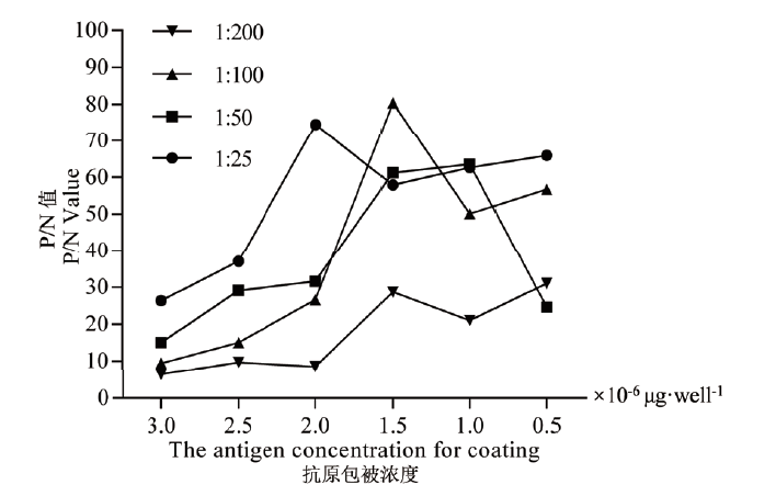
图1 抗原包被量及不同血清稀释度P/N值方正滴定结果
Fig. 1 Checkerboard titration results of P/N values for the epitope antigen coating amount and different serum dilution
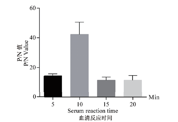
图2 血清反应时间优化结果
Fig. 2 Optimized results of serum reaction time
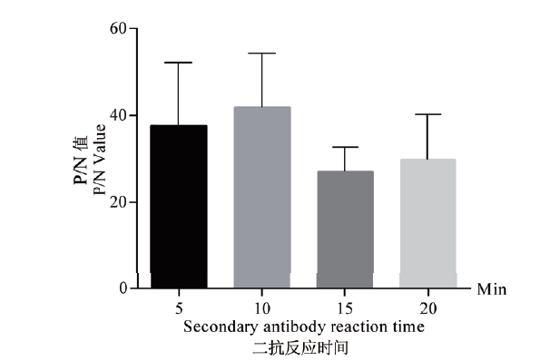
图3 酶标二抗反应时间优化结果
Fig. 3 Optimized results of secondary antibody reaction time

图4 H-CLIA临界值、诊断灵敏性和特异性分析结果 A:H-CLIA ROC曲线分析;B:H-CLIA TG-ROC曲线分析;C:H-CLIA 交互式点状图分析,“0”代表阴性血清样本,“1”代表阳性血清样本
Fig. 4 Results of cut-off values, diagnostic sensitivity, and specificity for H-CLIA A: H-CLIA ROC analysis curve. B: H-CLIA TG-ROC analysis curve. C: Interactive dot diagram of H-CLIA; 0: negative serum samples; 1: positive serum samples
| 标准Criterion | 敏感性Sensitivity | 95%置信区间95% CI | 特异性Specificity | 95%置信区间95% CI | 阳性似然比+LR | 阴性似然比-LR |
|---|---|---|---|---|---|---|
| > 8.72 | 96.34 | 93.7-98.1 | 89.32 | 81.7-94.5 | 9.02 | 0.041 |
| > 8.88 | 96.34 | 93.7-98.1 | 90.29 | 82.9-95.2 | 9.92 | 0.041 |
| > 9.06 | 96.34 | 93.7-98.1 | 91.26 | 84.1-95.9 | 11.03 | 0.040 |
| > 9.4 | 96.04 | 93.3-97.9 | 91.26 | 84.1-95.9 | 10.99 | 0.043 |
| > 9.52 | 95.73 | 92.9-97.6 | 91.26 | 84.1-95.9 | 10.96 | 0.047 |
| > 9.56 | 95.43 | 92.6-97.4 | 91.26 | 84.1-95.9 | 10.92 | 0.050 |
| > 9.77 | 95.43 | 92.6-97.4 | 92.23 | 85.3-96.6 | 12.29 | 0.050 |
| > 9.93 | 95.12 | 92.2-97.2 | 92.23 | 85.3-96.6 | 12.25 | 0.053 |
| > 10.07 | 94.82 | 91.8-97.0 | 92.23 | 85.3-96.6 | 12.21 | 0.056 |
| > 10.41 | 94.51 | 91.5-96.7 | 92.23 | 85.3-96.6 | 12.17 | 0.059 |
| > 10.45 | 94.21 | 91.1-96.5 | 92.23 | 85.3-96.6 | 12.13 | 0.063 |
| > 10.77 | 93.90 | 90.7-96.2 | 92.23 | 85.3-96.6 | 12.09 | 0.066 |
| > 10.92 | 93.60 | 90.4-96.0 | 92.23 | 85.3-96.6 | 12.05 | 0.069 |
表2 不同临界值对应的灵敏性及特异性
Table 2 Diagnosis sensitivity and specificity corresponding to different cut-off values
| 标准Criterion | 敏感性Sensitivity | 95%置信区间95% CI | 特异性Specificity | 95%置信区间95% CI | 阳性似然比+LR | 阴性似然比-LR |
|---|---|---|---|---|---|---|
| > 8.72 | 96.34 | 93.7-98.1 | 89.32 | 81.7-94.5 | 9.02 | 0.041 |
| > 8.88 | 96.34 | 93.7-98.1 | 90.29 | 82.9-95.2 | 9.92 | 0.041 |
| > 9.06 | 96.34 | 93.7-98.1 | 91.26 | 84.1-95.9 | 11.03 | 0.040 |
| > 9.4 | 96.04 | 93.3-97.9 | 91.26 | 84.1-95.9 | 10.99 | 0.043 |
| > 9.52 | 95.73 | 92.9-97.6 | 91.26 | 84.1-95.9 | 10.96 | 0.047 |
| > 9.56 | 95.43 | 92.6-97.4 | 91.26 | 84.1-95.9 | 10.92 | 0.050 |
| > 9.77 | 95.43 | 92.6-97.4 | 92.23 | 85.3-96.6 | 12.29 | 0.050 |
| > 9.93 | 95.12 | 92.2-97.2 | 92.23 | 85.3-96.6 | 12.25 | 0.053 |
| > 10.07 | 94.82 | 91.8-97.0 | 92.23 | 85.3-96.6 | 12.21 | 0.056 |
| > 10.41 | 94.51 | 91.5-96.7 | 92.23 | 85.3-96.6 | 12.17 | 0.059 |
| > 10.45 | 94.21 | 91.1-96.5 | 92.23 | 85.3-96.6 | 12.13 | 0.063 |
| > 10.77 | 93.90 | 90.7-96.2 | 92.23 | 85.3-96.6 | 12.09 | 0.066 |
| > 10.92 | 93.60 | 90.4-96.0 | 92.23 | 85.3-96.6 | 12.05 | 0.069 |
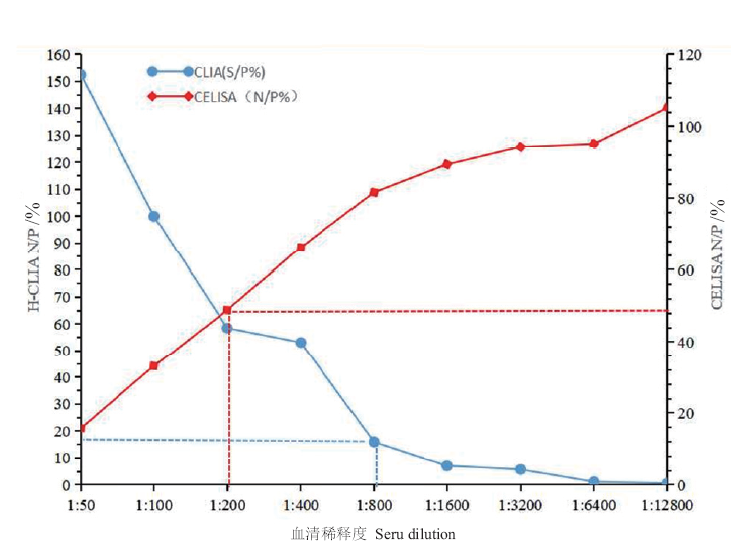
图5 H-CLIA和cELISA的灵敏性比对
Fig. 5 Comparison of the analytical sensitivity between H-CLIA and cELISA
| 血清编号 Serum No. | 检测样品S/P值 S/P value of a detection sample/% | 结果判定 Result determination | ||
|---|---|---|---|---|
| A | B | C | ||
| PPRV(+) | 18.17 | 22.13 | 13.46 | + |
| PPRV(-) | 0.27 | 0.33 | 0.28 | - |
| FMDV(+) | 7.74 | 9.10 | 5.85 | - |
| SPV(+) | 5.78 | 6.37 | 9.45 | - |
| BTV(+) | 3.31 | 6.69 | 8.48 | - |
| MCCP(+) | 5.74 | 4.72 | 2.04 | - |
表3 特异性检测
Table 3 Specificity detection
| 血清编号 Serum No. | 检测样品S/P值 S/P value of a detection sample/% | 结果判定 Result determination | ||
|---|---|---|---|---|
| A | B | C | ||
| PPRV(+) | 18.17 | 22.13 | 13.46 | + |
| PPRV(-) | 0.27 | 0.33 | 0.28 | - |
| FMDV(+) | 7.74 | 9.10 | 5.85 | - |
| SPV(+) | 5.78 | 6.37 | 9.45 | - |
| BTV(+) | 3.31 | 6.69 | 8.48 | - |
| MCCP(+) | 5.74 | 4.72 | 2.04 | - |
| 血清编号 Serum No. | 同一批次S/P值S/P value of same batch/% | 平均值 Average value | 标准差 Standard deviation | 变异系数 Coefficient of variation CV/% | ||
|---|---|---|---|---|---|---|
| A | B | C | ||||
| 1 | 7.33 | 8.03 | 8.48 | 7.95 | 0.47 | 5.96 |
| 2 | 8.09 | 8.80 | 7.71 | 8.20 | 0.45 | 5.51 |
| 3 | 4.390 | 5.26 | 5.45 | 5.03 | 0.46 | 9.17 |
| 4 | 8.040 | 7.77 | 8.88 | 8.23 | 0.47 | 5.74 |
| 5 | 19.79 | 22.89 | 22.3 | 21.66 | 1.34 | 6.21 |
| 6 | 18.48 | 19.68 | 22.33 | 20.16 | 1.61 | 7.98 |
| 7 | 14.01 | 11.98 | 12.65 | 12.88 | 0.84 | 6.56 |
| 8 | 40.98 | 43.32 | 40.11 | 41.47 | 1.36 | 3.27 |
表4 批内重复性检测结果
Table 4 Results of intra-run repeatability experiment
| 血清编号 Serum No. | 同一批次S/P值S/P value of same batch/% | 平均值 Average value | 标准差 Standard deviation | 变异系数 Coefficient of variation CV/% | ||
|---|---|---|---|---|---|---|
| A | B | C | ||||
| 1 | 7.33 | 8.03 | 8.48 | 7.95 | 0.47 | 5.96 |
| 2 | 8.09 | 8.80 | 7.71 | 8.20 | 0.45 | 5.51 |
| 3 | 4.390 | 5.26 | 5.45 | 5.03 | 0.46 | 9.17 |
| 4 | 8.040 | 7.77 | 8.88 | 8.23 | 0.47 | 5.74 |
| 5 | 19.79 | 22.89 | 22.3 | 21.66 | 1.34 | 6.21 |
| 6 | 18.48 | 19.68 | 22.33 | 20.16 | 1.61 | 7.98 |
| 7 | 14.01 | 11.98 | 12.65 | 12.88 | 0.84 | 6.56 |
| 8 | 40.98 | 43.32 | 40.11 | 41.47 | 1.36 | 3.27 |
| 血清编号Serum No. | 不同批次S/P值 S/P values of different batches/% | 平均值 Average value | 标准差 Standard deviation | 变异系数 Coefficient of variation CV/% | ||
|---|---|---|---|---|---|---|
| 1 | 2 | 3 | ||||
| 1 | 7.55 | 7.33 | 6.29 | 7.06 | 0.55 | 7.79 |
| 2 | 5.59 | 5.68 | 6.65 | 5.97 | 0.48 | 8.03 |
| 3 | 1.92 | 1.97 | 2.01 | 1.97 | 0.04 | 1.87 |
| 4 | 8.29 | 7.86 | 6.67 | 7.61 | 0.69 | 9.01 |
| 5 | 32.36 | 32.06 | 26.89 | 30.44 | 2.51 | 8.25 |
| 6 | 22.82 | 18.18 | 19.79 | 20.26 | 1.92 | 9.49 |
| 7 | 45.11 | 53.18 | 51.55 | 49.95 | 3.48 | 6.98 |
| 8 | 34.97 | 35.87 | 35.27 | 35.37 | 0.37 | 1.06 |
表5 批间重复性检测结果
Table 5 Results of inter-run repeatability experiment
| 血清编号Serum No. | 不同批次S/P值 S/P values of different batches/% | 平均值 Average value | 标准差 Standard deviation | 变异系数 Coefficient of variation CV/% | ||
|---|---|---|---|---|---|---|
| 1 | 2 | 3 | ||||
| 1 | 7.55 | 7.33 | 6.29 | 7.06 | 0.55 | 7.79 |
| 2 | 5.59 | 5.68 | 6.65 | 5.97 | 0.48 | 8.03 |
| 3 | 1.92 | 1.97 | 2.01 | 1.97 | 0.04 | 1.87 |
| 4 | 8.29 | 7.86 | 6.67 | 7.61 | 0.69 | 9.01 |
| 5 | 32.36 | 32.06 | 26.89 | 30.44 | 2.51 | 8.25 |
| 6 | 22.82 | 18.18 | 19.79 | 20.26 | 1.92 | 9.49 |
| 7 | 45.11 | 53.18 | 51.55 | 49.95 | 3.48 | 6.98 |
| 8 | 34.97 | 35.87 | 35.27 | 35.37 | 0.37 | 1.06 |
| 检测方法Detecting method | H-CLIA | 阳性 Positive | 阴性 Negative | 合计 Sum |
|---|---|---|---|---|
| cELISA | 阳性Positive | 77 | 0 | 77 |
| 阴性Negative | 14 | 156 | 170 | |
| 合计Sum | 91 | 156 | 247 | |
| 符合率Coincidence rate/% | - | - | 94.33 |
表6 比对试验相对符合率统计
Table 6 Coincidence rate of comparison test
| 检测方法Detecting method | H-CLIA | 阳性 Positive | 阴性 Negative | 合计 Sum |
|---|---|---|---|---|
| cELISA | 阳性Positive | 77 | 0 | 77 |
| 阴性Negative | 14 | 156 | 170 | |
| 合计Sum | 91 | 156 | 247 | |
| 符合率Coincidence rate/% | - | - | 94.33 |
| [1] |
Bailey D, Banyard A, Dash P, et al. Full genome sequence of peste des petits ruminants virus, a member of the Morbillivirus genus[J]. Virus Res, 2005, 110(1-2): 119-124.
pmid: 15845262 |
| [2] |
Balamurugan V, Hemadri D, Gajendragad MR, et al. Diagnosis and control of peste des petits ruminants: a comprehensive review[J]. Virusdisease, 2014, 25(1): 39-56.
doi: 10.1007/s13337-013-0188-2 pmid: 24426309 |
| [3] |
Balamurugan V, Sen A, Venkatesan G, et al. Sequence and phylogenetic analyses of the structural genes of virulent isolates and vaccine strains of peste des petits ruminants virus from India[J]. Transbound Emerg Dis, 2010, 57(5): 352-364.
doi: 10.1111/j.1865-1682.2010.01156.x pmid: 20642492 |
| [4] |
Kwiatek O, Ali YH, Saeed IK, et al. Asian lineage of peste des petits ruminants virus, Africa[J]. Emerg Infect Dis, 2011, 17(7): 1223-1231.
doi: 10.3201/eid1707.101216 pmid: 21762576 |
| [5] |
Banyard AC, Parida S, Batten C, et al. Global distribution of peste des petits ruminants virus and prospects for improved diagnosis and control[J]. J Gen Virol, 2010, 91(Pt 12): 2885-2897.
doi: 10.1099/vir.0.025841-0 pmid: 20844089 |
| [6] |
Gibbs EP, Taylor WP, Lawman MJ, et al. Classification of peste des petits ruminants virus as the fourth member of the genus Morbillivirus[J]. Intervirology, 1979, 11(5): 268-274.
pmid: 457363 |
| [7] |
Dou YX, Liang ZX, Prajapati M, et al. Expanding diversity of susceptible hosts in peste des petits ruminants virus infection and its potential mechanism beyond[J]. Front Vet Sci, 2020, 7: 66.
doi: 10.3389/fvets.2020.00066 pmid: 32181263 |
| [8] |
Albayrak H, Gür S. A serologic investigation for Peste des petits ruminants infection in sheep, cattle and camels(Camelus dromedarius)in Aydin province, West Anatolia[J]. Trop Anim Health Prod, 2010, 42(2): 151-153.
doi: 10.1007/s11250-009-9400-1 pmid: 19554466 |
| [9] |
Intisar KS, Ali YH, Haj MA, et al. Peste des petits ruminants infection in domestic ruminants in Sudan[J]. Trop Anim Health Prod, 2017, 49(4): 747-754.
doi: 10.1007/s11250-017-1254-3 pmid: 28321790 |
| [10] |
Maillard JC, Van KP, Nguyen T, et al. Examples of probable host-pathogen co-adaptation/co-evolution in isolated farmed animal populations in the mountainous regions of North Vietnam[J]. Ann N Y Acad Sci, 2008, 1149: 259-262.
doi: 10.1196/nyas.2008.1149.issue-1 URL |
| [11] |
Zakian A, Nouri M, Kahroba H, et al. The first report of peste des petits ruminants(PPR)in camels(Camelus dromedarius)in Iran[J]. Trop Anim Health Prod, 2016, 48(6): 1215-1219.
doi: 10.1007/s11250-016-1078-6 pmid: 27155951 |
| [12] |
Ratta B, Pokhriyal M, Singh SK, et al. Detection of peste des petits ruminants virus(PPRV)genome from nasal swabs of dogs[J]. Curr Microbiol, 2016, 73(1): 99-103.
doi: 10.1007/s00284-016-1030-z URL |
| [13] |
Furley CW, Taylor WP, Obi TU. An outbreak of peste des petits ruminants in a zoological collection[J]. Vet Rec, 1987, 121(19): 443-447.
doi: 10.1136/vr.121.19.443 URL |
| [14] |
Schulz C, Fast C, Schlottau K, et al. Neglected hosts of small ruminant Morbillivirus[J]. Emerg Infect Dis, 2018, 24(12): 2334-2337.
doi: 10.3201/eid2412.180507 URL |
| [15] |
Liu F, Li J, Li L, et al. Peste des petits ruminants in China since its first outbreak in 2007: a 10-year review[J]. Transbound Emerg Dis, 2018, 65(3): 638-648.
doi: 10.1111/tbed.12808 pmid: 29322642 |
| [16] |
Santhamani R, Singh RP, Njeumi F. Peste des petits ruminants diagnosis and diagnostic tools at a glance: perspectives on global control and eradication[J]. Arch Virol, 2016, 161(11): 2953-2967.
doi: 10.1007/s00705-016-3009-2 pmid: 27522587 |
| [17] | 孙雨, 赵柏林, 王晓英, 等. 小反刍兽疫病毒N蛋白原核表达与间接ELISA检测方法的建立[J]. 动物医学进展, 2017, 38(1): 6-10. |
| Sun Y, Zhao BL, Wang XY, et al. Prokaryotic expression of N protein of peste des petits ruminants virus and development of indirect ELISA[J]. Prog Vet Med, 2017, 38(1): 6-10. | |
| [18] |
Taylor W. The global eradication of peste des petits ruminants(PPR)within 15 years—is this a pipe dream?[J]. Trop Anim Health Prod, 2016, 48(3): 559-567.
doi: 10.1007/s11250-016-0993-x URL |
| [19] |
Vongpunsawad S, Oezgun N, Braun W, et al. Selectively receptor-blind measles viruses: identification of residues necessary for SLAM- or CD46-induced fusion and their localization on a new hemagglutinin structural model[J]. J Virol, 2004, 78(1): 302-313.
pmid: 14671112 |
| [20] |
Yoneda M, Bandyopadhyay SK, Shiotani M, et al. Rinderpest virus H protein: role in determining host range in rabbits[J]. J Gen Virol, 2002, 83(Pt 6): 1457-1463.
doi: 10.1099/0022-1317-83-6-1457 pmid: 12029161 |
| [21] | 梁忠祥. 小反刍兽疫多表位疫苗抗原的设计及其免疫效力评价[D]. 北京: 中国农业科学院, 2017. |
| Liang ZX. Design and evaluation of the multi-epitope antigens against peste des petits ruminants[D]. Beijing: Chinese Academy of Agricultural Sciences, 2017. | |
| [22] | 钱榜, 李彦敏, 朱学亮, 等. 基于H蛋白表位合成肽的小反刍兽疫病毒抗体间接ELISA检测方法的建立[J]. 畜牧兽医学报, 2021, 52(1): 144-153. |
| Qian B, Li YM, Zhu XL, et al. Establishment of an iELISA method for detection of antibody againist peste des petits ruminants virus based on H protein epitope peptide[J]. Acta Vet Zootechnica Sin, 2021, 52(1): 144-153. | |
| [23] |
Frey A, di Canzio J, Zurakowski D. A statistically defined endpoint titer determination method for immunoassays[J]. J Immunol Methods, 1998, 221(1/2): 35-41.
doi: 10.1016/S0022-1759(98)00170-7 URL |
| [24] |
Liu ZZ, Zhao FR, Gao SD, et al. Development of a chemiluminescence immunoassay using recombinant non-structural epitope-based proteins to accurately differentiate foot-and-mouth disease virus-infected and vaccinated bovines[J]. Transbound Emerg Dis, 2018, 65(2): 338-344.
doi: 10.1111/tbed.12811 pmid: 29341485 |
| [25] |
Pruvot M, Fine AE, Hollinger C, et al. Outbreak of peste des petits ruminants among critically endangered Mongolian Saiga and other wild ungulates, Mongolia, 2016-2017[J]. Emerg Infect Dis, 2020, 26(1): 51-62.
doi: 10.3201/eid2601.181998 URL |
| [26] | Mdetele DP, Komba E, Seth MD, et al. Review of peste des petits ruminants occurrence and spread in Tanzania[J]. Animals(Basel), 2021, 11(6): 1698. |
| [27] |
Aziz-Ul-Rahman, Wensman JJ, Abubakar M, et al. Peste des petits ruminants in wild ungulates[J]. Trop Anim Health Prod, 2018, 50(8): 1815-1819.
doi: 10.1007/s11250-018-1623-6 pmid: 29881925 |
| [28] |
Rajak KK, Sreenivasa BP, Hosamani M, et al. Experimental studies on immunosuppressive effects of peste des petits ruminants(PPR)virus in goats[J]. Comp Immunol Microbiol Infect Dis, 2005, 28(4): 287-296.
doi: 10.1016/j.cimid.2005.08.002 URL |
| [29] |
Choi KS, Nah JJ, Ko YJ, et al. Localization of antigenic sites at the amino-Terminus of rinderpest virus N protein using deleted N mutants and monoclonal antibody[J]. J Vet Sci, 2003, 4(2): 167-173.
doi: 10.4142/jvs.2003.4.2.167 URL |
| [30] |
Roda A, Guarigli M, Michelini E, et al. Analytical bioluminescence and chemiluminescence[J]. Anal Chem, 2003, 75(21): 463A-470A.
pmid: 14619853 |
| [31] | 胥传来, 彭池方, 郝凯, 金征宇, 王武康. 化学发光酶免疫方法检测克伦特罗残留[J]. 分析化学, 2005, 33(5): 699-702. |
| Xu CL, Peng CF, Hao K, et al. Determination of clenbuterol residual by chemiluminescent enzyme immunossay[J]. Chin J Anal Chem, 2005, 33(5): 699-702. | |
| [32] | 廖晓阳, 金成勇. 如何在诊断性试验研究中正确应用有关测量指标[J]. 华西医学, 2000, 15(2): 146-147. |
| Liao XY, Jin CY. How to effectively using diagnostic measurement to study diagnostic test[J]. West China Med J, 2000, 15(2): 146-147. | |
| [33] | 梁万年, 王存亮. 如何应用四格表评估诊断试验的价值[J]. 中国全科医学, 2005, 8(4): 288-289. |
| Liang WN, Wang CL. How to using a four-grid table to evaluate the value of a diagnostic test[J]. Chin Gen Pract, 2005, 8(4): 288-289. | |
| [34] |
Renukaradhya GJ, Sinnathamby G, Seth S, et al. Mapping of B-cell epitopic sites and delineation of functional domains on the hemagglutinin-neuraminidase protein of peste des petits ruminants virus[J]. Virus Res, 2002, 90(1-2): 171-185.
pmid: 12457972 |
| [35] |
Libeau G, Diallo A, Calvez D, et al. A competitive ELISA using anti-N monoclonal antibodies for specific detection of rinderpest antibodies in cattle and small ruminants[J]. Vet Microbiol, 1992, 31(2/3): 147-160.
doi: 10.1016/0378-1135(92)90073-3 URL |
| [36] |
Singh RP, Sreenivasa BP, Dhar P, et al. A sandwich-ELISA for the diagnosis of Peste des petits ruminants(PPR)infection in small ruminants using anti-nucleocapsid protein monoclonal antibody[J]. Arch Virol, 2004, 149(11): 2155-2170.
pmid: 15503204 |
| [37] | 栾志舫, 陈妍, 吴锦艳, 等. 小反刍兽疫病毒H蛋白的表达及间接ELISA方法的建立[J]. 中国兽医学报, 2016, 36(7): 1109-1114. |
| Luan ZF, Chen Y, Wu JY, et al. Prokaryotic expression of H protein of peste des petits ruminants virus and development of indirect ELISA[J]. Chin J Vet Sci, 2016, 36(7): 1109-1114. |
| [1] | 李丹, 杜梦潭, 修明霞, 刘兴健, 张志芳, 李轶女. 羊α干扰素在家蚕中的表达及抗小反刍兽疫病毒活性测定[J]. 生物技术通报, 2022, 38(1): 187-193. |
| [2] | 郭磊周, 韩佳慧, 唐殷, 李江, 黄程, 代其林, 王劲, 平淑珍, 江世杰. DrwH类信号肽序列对其抗氧化功能的影响[J]. 生物技术通报, 2019, 35(5): 125-132. |
| [3] | 邓瑞雪, 蒙学莲, 曾巧英, 才学鹏. 小反刍兽疫病毒F基因缺失体的克隆、表达及鉴定[J]. 生物技术通报, 2016, 32(5): 234-239. |
| [4] | 李江涛;殷相平;张金卫;丁农;柳纪省;. 狂犬病病毒CVS株糖蛋白、核蛋白生物信息学分析[J]. , 2010, 0(07): 179-184. |
| [5] | 李任峰;田香勤;何启盖;王自良;赵坤;李学斌;王三虎;. 猪胸膜肺炎放线杆菌ApxIIA基因的序列分析及其B细胞表位预测[J]. , 2009, 0(07): 104-108. |
| [6] | 金定恩;张力青;姚艳丰;韩丽;梁爱心;滑国华;杨利国;. 猪肌生成抑制素基因去信号肽蛋白二级构预测和B细胞表位分析[J]. , 2008, 0(S1): 264-270. |
| [7] | 张昱;王永录;张永光;潘丽;方玉珍;刘力宽;蒋守田;吕建亮;张中旺;张淑刚;李正丰;杜进鑫;. 口蹄疫病毒株AF72 VP1的结构构建与B细胞表位预测[J]. , 2008, 0(06): 158-163. |
| 阅读次数 | ||||||
|
全文 |
|
|||||
|
摘要 |
|
|||||