







Biotechnology Bulletin ›› 2026, Vol. 42 ›› Issue (10): 328-338.doi: 10.13560/j.cnki.biotech.bull.1985.2025-1397
YANG Tai-ran1,2,3,4, ZHENG Xin-qi1,2,3,4, HU Chao2,3,4,5, AN Ting-ting2,3,4,5, WEN Long2,3,4,5, LI You-li2,3,4,5, CHEN Zheng-hong2,3,5, CUI Gu-zhen2,3,5, LI Bo-yan1( ), TIAN Yi-ming2,3,4,5()
Received:2025-12-22
Online:2026-10-26
Published:2026-10-15
Contact:
LI Bo-yan, TIAN Yi-ming
E-mail:boyan.li@gmc.edu.cn;tianym@gmc.edu.cn
YANG Tai-ran, ZHENG Xin-qi, HU Chao, AN Ting-ting, WEN Long, LI You-li, CHEN Zheng-hong, CUI Gu-zhen, LI Bo-yan, TIAN Yi-ming. Construction and Functional Characterization of Stable Cell Lines Expressing Nucleocapsid Protein of Porcine Deltacoronavirus[J]. Biotechnology Bulletin, 2026, 42(10): 328-338.
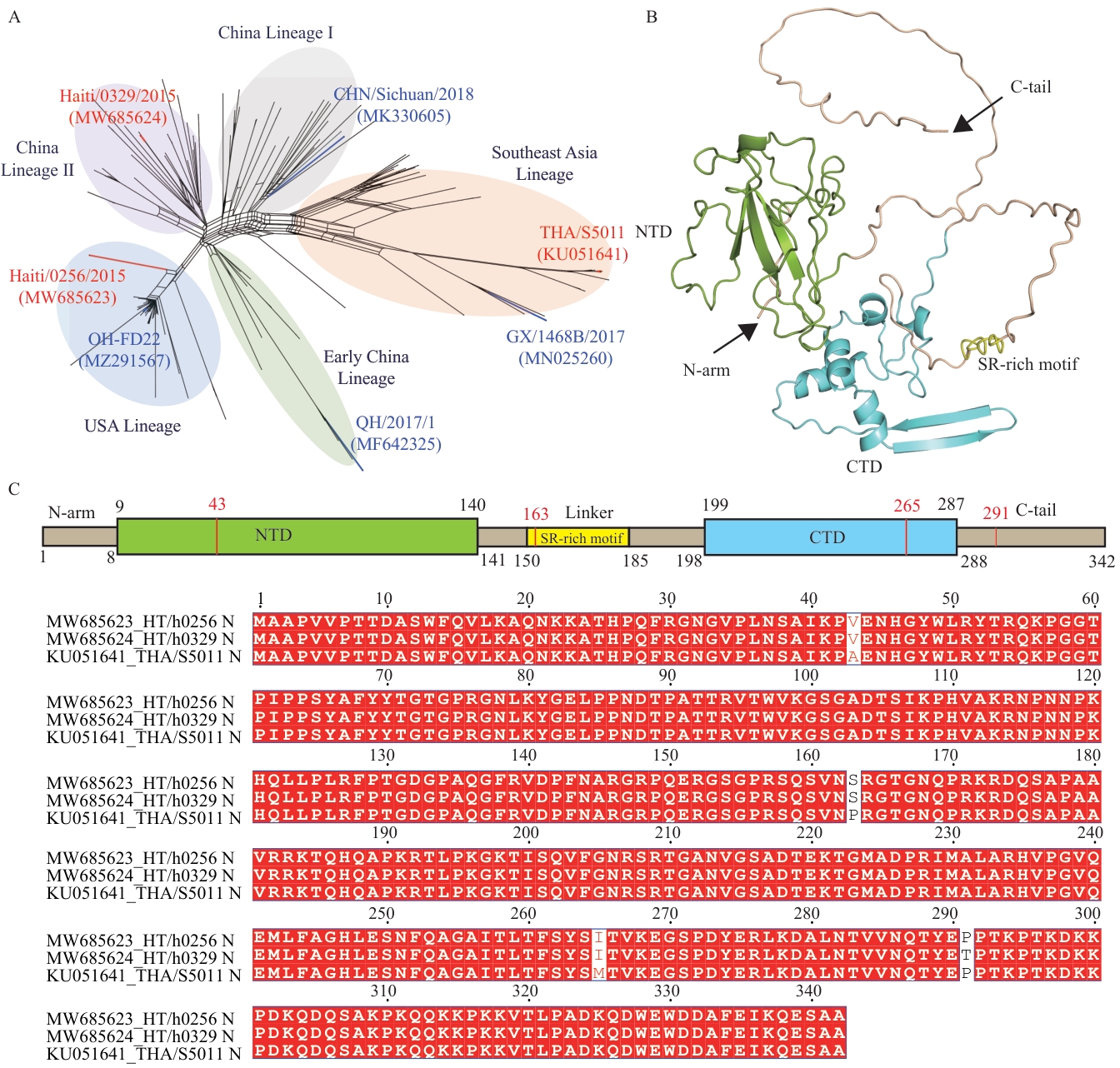
Fig. 1 Functional domain and amino acid levels analysis of N protein from three PDCoV lineagesA: Phylogenetic genotyping based on 194 complete-genome sequences of PDCoV (red indicates representative strains of PDCoV from three lineages). B: Schematic diagram of PDCoV N protein domain (Green: NTD. Cyan: CTD. Yellow: SR-rich motif). C: Amino acid sequence alignment of N proteins from three PDCoV lineages
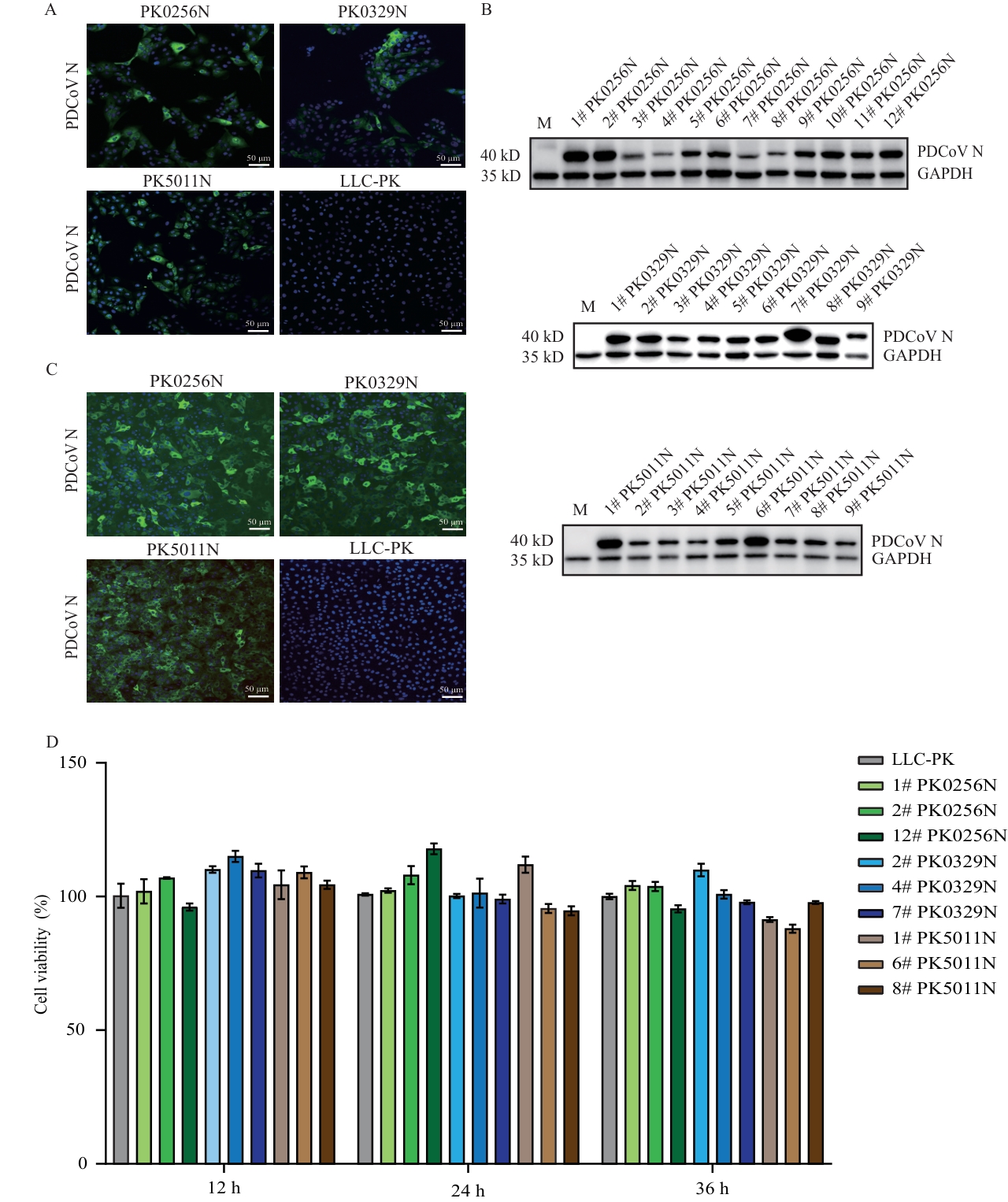
Fig. 2 Construction and identification of stable cell lines expressing the N protein from three PDCoV lineagesA: Indirect immunofluorescence assay for detecting the expression and localization of PDCoV N protein from three lineages in polyclonal cell lines (scale bar: 50 μm). B: Western blot analysis of N protein expression levels in monoclonal stable cell lines of three PDCoV lineages (M: LLC-PK control cells). C: Indirect immunofluorescence assay for detecting the expression of N protein in monoclonal stable cell lines of three PDCoV lineages (scale bar: 50 μm). D: CCK-8 assay for evaluating the proliferative activity of stable cell lines of three PDCoV lineages; the ordinate represents cell viability, and the abscissa represents detection time points. *P<0.05, **P<0.01, ***P<0.001, ****P<0.000 1. No asterisk indicates no statistically significant difference between groups. The same below
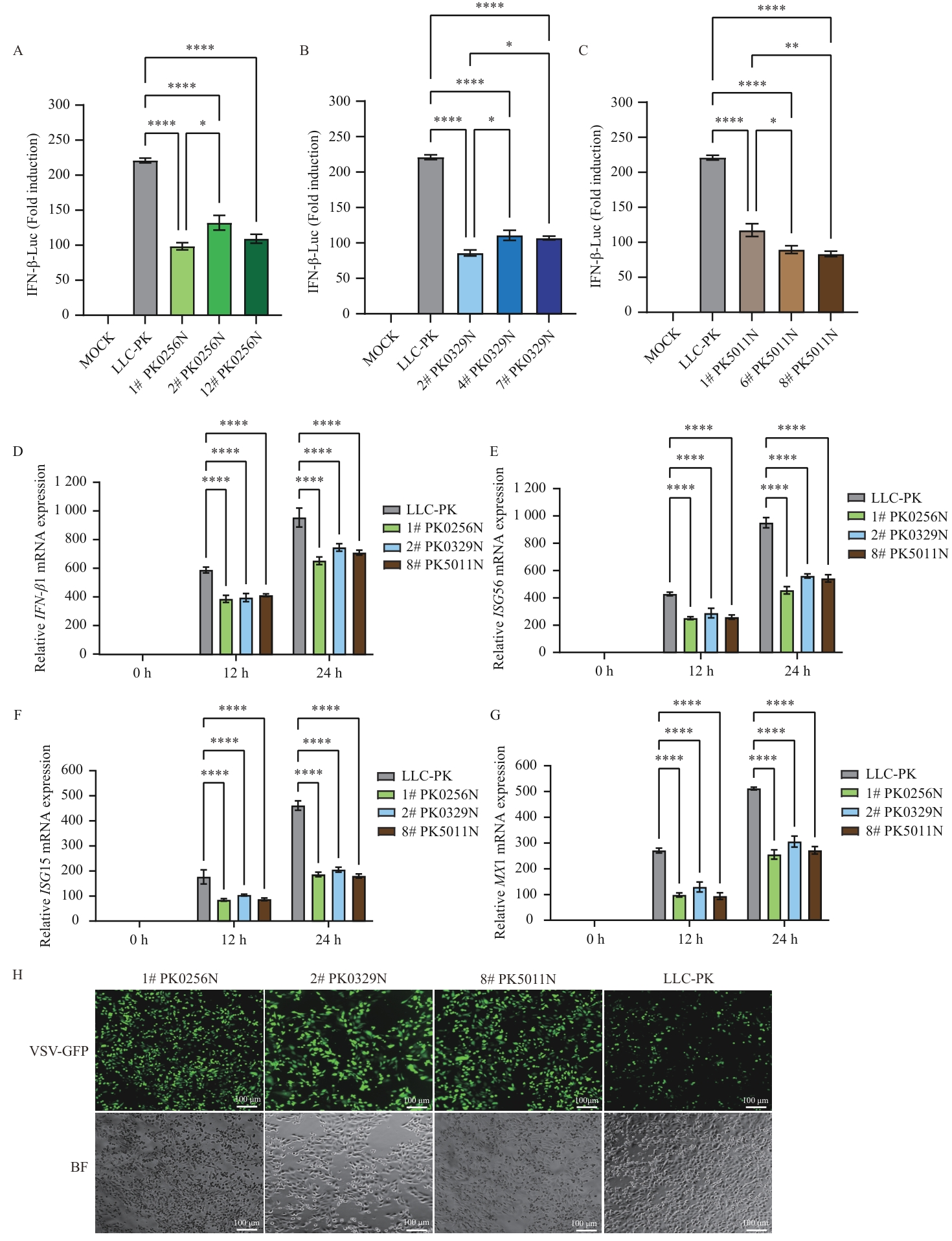
Fig. 3 PDCoV N protein suppresses IFN-β activation and downstream stimulated gene expressionA‒C: Analysis of the effect of N proteins from three PDCoV lineages on SeV-induced IFN-β promoter activity using a dual-luciferase reporter assay system (MOCK: Mock control). D‒G: Detection of the relative mRNA expression levels of SeV-induced IFN-β1 (D), ISG56 (E), ISG15 (F) and MX1 (G) genes in stable cell lines of different PDCoV lineages by RT-qPCR. H: Fluorescence microscopy observation of the effect of PDCoV N protein on VSV-GFP replication (green fluorescence intensity indicates VSV replication level. scale bar: 100 μm)
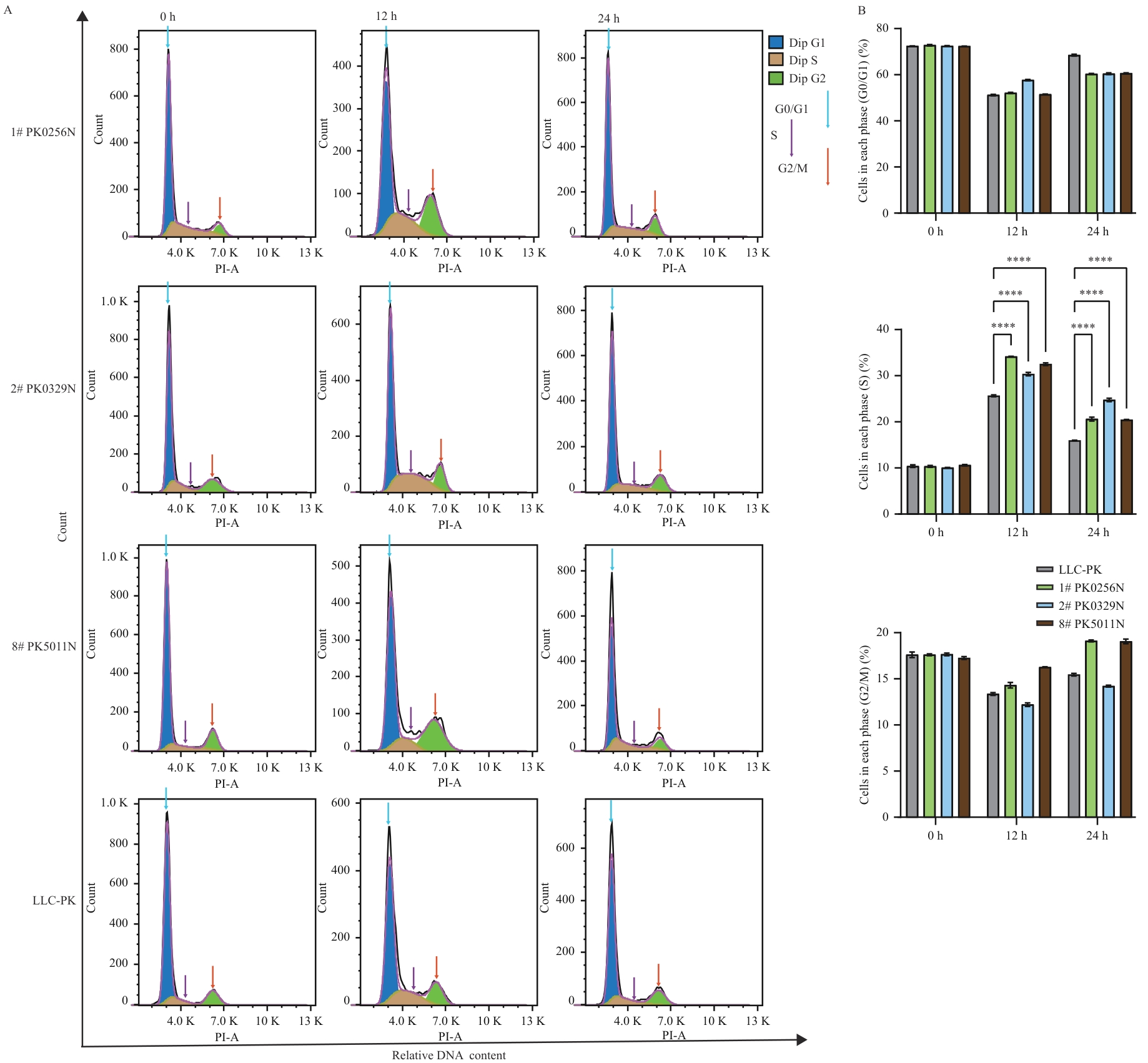
Fig. 4 PDCoV N protein induces S-phase arrest in LLC-PK cellsA: Representative cell cycle profiles of wild-type LLC-PK cells and PDCoV N protein stably transfected cell lines detected by flow cytometry at different time points (0, 12 and 24 h) after serum starvation synchronization (Blue: G0/G1 phase. Brown: S phase. Green: G2/M phase). B: Statistical results of the proportions of cells in G0/G1, S and G2/M phases at different time points
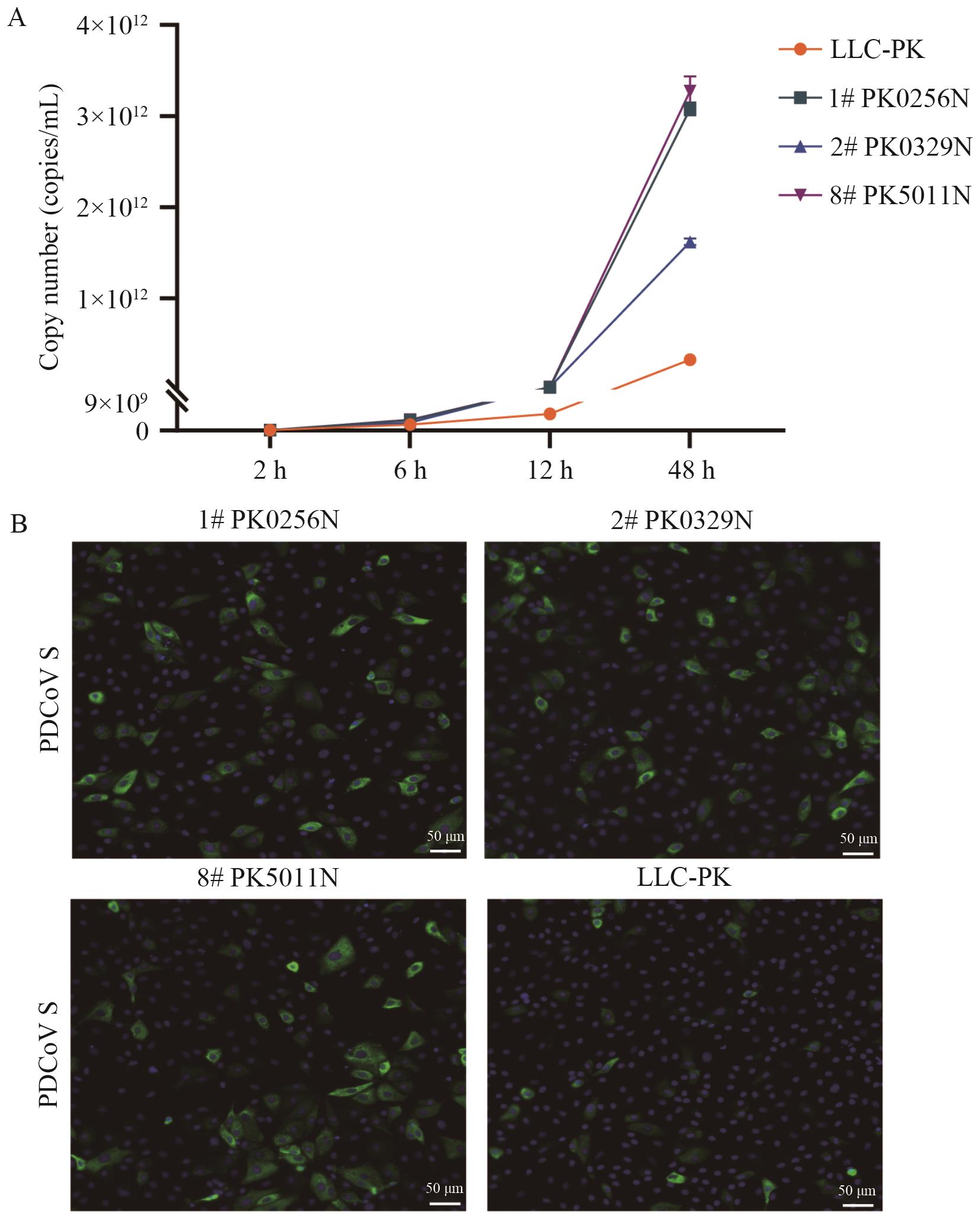
Fig. 5 PDCoV N protein enhances PDCoV replication and proliferationA: Replication kinetics of PDCoV in supernatants of N protein stable cell lines from three lineages using absolute quantitative qPCR. B: Immunofluorescence detection of PDCoV proliferation in stable cell lines expressing the N protein using an anti-spike monoclonal antibody (scale bar: 50 μm)
| [1] | Cui J, Li F, Shi ZL. Origin and evolution of pathogenic coronaviruses [J]. Nat Rev Microbiol, 2019, 17(3): 181-192. |
| [2] | Ye ZW, Yuan SF, Yuen KS, et al. Zoonotic origins of human coronaviruses [J]. Int J Biol Sci, 2020, 16(10): 1686-1697. |
| [3] | Ruiz-Aravena M, McKee C, Gamble A, et al. Ecology, evolution and spillover of coronaviruses from bats [J]. Nat Rev Microbiol, 2022, 20(5): 299-314. |
| [4] | Miranda C, Silva V, Igrejas G, et al. Genomic evolution of the human and animal coronavirus diseases [J]. Mol Biol Rep, 2021, 48(9): 6645-6653. |
| [5] | Zhang JQ. Porcine deltacoronavirus: Overview of infection dynamics, diagnostic methods, prevalence and genetic evolution [J]. Virus Res, 2016, 226: 71-84. |
| [6] | Wang QH, Vlasova AN, Kenney SP, et al. Emerging and re-emerging coronaviruses in pigs [J]. Curr Opin Virol, 2019, 34: 39-49. |
| [7] | Shan XT, Li R, Ma X, et al. Epidemiology, pathogenesis, immune evasion mechanism and vaccine development of porcine Deltacoronavirus [J]. Funct Integr Genomics, 2024, 24(3): 79. |
| [8] | He WT, Ji X, He W, et al. Genomic epidemiology, evolution, and transmission dynamics of porcine deltacoronavirus [J]. Mol Biol Evol, 2020, 37(9): 2641-2654. |
| [9] | Tang P, Cui EH, Song YH, et al. Porcine deltacoronavirus and its prevalence in China: a review of epidemiology, evolution, and vaccine development [J]. Arch Virol, 2021, 166(11): 2975-2988. |
| [10] | Bahoussi AN, Wang PH, Shah PT, et al. Evolutionary plasticity of zoonotic porcine Deltacoronavirus (PDCoV): genetic characteristics and geographic distribution [J]. BMC Vet Res, 2022, 18: 444. |
| [11] | Jung K, Hu H, Saif LJ. Porcine deltacoronavirus infection: Etiology, cell culture for virus isolation and propagation, molecular epidemiology and pathogenesis [J]. Virus Res, 2016, 226: 50-59. |
| [12] | Vlasova AN, Kenney SP, Jung K, et al. Deltacoronavirus evolution and transmission: current scenario and evolutionary perspectives [J]. Front Vet Sci, 2021, 7: 626785. |
| [13] | Tian YY, Sun JQ, Hou XH, et al. Cross-species recognition of two porcine coronaviruses to their cellular receptor aminopeptidase N of dogs and seven other species [J]. PLoS Pathog, 2025, 21(1): e1012836. |
| [14] | Lednicky JA, Tagliamonte MS, White SK, et al. Independent infections of porcine deltacoronavirus among Haitian children [J]. Nature, 2021, 600(7887): 133-137. |
| [15] | Su S, Wong G, Shi WF, et al. Epidemiology, genetic recombination, and pathogenesis of coronaviruses [J]. Trends Microbiol, 2016, 24(6): 490-502. |
| [16] | Ye QZ, West AMV, Silletti S, et al. Architecture and self-assembly of the SARS-CoV-2 nucleocapsid protein [J]. Protein Sci, 2020, 29(9): 1890-1901. |
| [17] | Chen H, Cui Y, Han XL, et al. Liquid-liquid phase separation by SARS-CoV-2 nucleocapsid protein and RNA [J]. Cell Res, 2020, 30(12): 1143-1145. |
| [18] | Cong YY, Ulasli M, Schepers H, et al. Nucleocapsid protein recruitment to replication-transcription complexes plays a crucial role in coronaviral life cycle [J]. J Virol, 2020, 94(4): e01925-e01919. |
| [19] | Mu JF, Fang YH, Yang Q, et al. SARS-CoV-2 N protein antagonizes type I interferon signaling by suppressing phosphorylation and nuclear translocation of STAT1 and STAT2 [J]. Cell Discov, 2020, 6: 65. |
| [20] | Pan P, Shen MM, Yu ZY, et al. SARS-CoV-2 N protein promotes NLRP3 inflammasome activation to induce hyperinflammation [J]. Nat Commun, 2021, 12: 4664. |
| [21] | Hu H, Jung K, Vlasova AN, et al. Isolation and characterization of porcine deltacoronavirus from pigs with diarrhea in the United States [J]. J Clin Microbiol, 2015, 53(5): 1537-1548. |
| [22] | Ji LK, Li SS, Zhu WX, et al. Porcine deltacoronavirus nucleocapsid protein suppressed IFN-β production by interfering porcine RIG-I dsRNA-binding and K63-linked polyubiquitination [J]. Front Immunol, 2019, 10: 1024. |
| [23] | Ji LK, Wang N, Ma JJ, et al. Porcine deltacoronavirus nucleocapsid protein species-specifically suppressed IRF7-induced type I interferon production via ubiquitin-proteasomal degradation pathway [J]. Vet Microbiol, 2020, 250: 108853. |
| [24] | Hu YT, Hao CL, Wang DH, et al. Porcine deltacoronavirus nucleocapsid protein antagonizes JAK-STAT signaling pathway by targeting STAT1 through KPNA2 degradation [J]. J Virol, 2024, 98(7): e00334-e00324. |
| [25] | Ji LK, Zhou LY, Wang Y, et al. FBXW8 suppresses PDCoV proliferation via the NPD52-dependent autophagic degradation of a viral nucleocapsid protein [J]. Front Immunol, 2024, 15: 1457255. |
| [26] | Yang XY, Kong N, Qin WZ, et al. PGAM5 degrades PDCoV N protein and activates type I interferon to antagonize viral replication [J]. J Virol, 2023, 97(11): e01470-e01423. |
| [27] | Wu XH, Lan SJ, Wang Y, et al. LDHB suppresses the PDCoV proliferation by targeting viral nucleocapsid protein for autophagic degradation [J]. Microbiol Spectr, 2025, 13(5): e02787-e02724. |
| [28] | Huson DH, Bryant D. Application of phylogenetic networks in evolutionary studies [J]. Mol Biol Evol, 2006, 23(2): 254-267. |
| [29] | Abramson J, Adler J, Dunger J, et al. Accurate structure prediction of biomolecular interactions with AlphaFold 3 [J]. Nature, 2024, 630(8016): 493-500. |
| [30] | Livak KJ, Schmittgen TD. Analysis of relative gene expression data using real-time quantitative PCR and the 2-ΔΔCT method [J]. Methods, 2001, 25(4): 402-408. |
| [31] | El-Maradny YA, Badawy MA, Mohamed KI, et al. Unraveling the role of the nucleocapsid protein in SARS-CoV-2 pathogenesis: From viral life cycle to vaccine development [J]. Int J Biol Macromol, 2024, 279: 135201. |
| [32] | Zhao YH, Sui LY, Wu P, et al. A dual-role of SARS-CoV-2 nucleocapsid protein in regulating innate immune response [J]. Sig Transduct Target Ther, 2021, 6: 331. |
| [33] | Liu JM, Liu MY, Yu XY, et al. SARS-CoV-2 nucleocapsid protein delays cell cycle in S-phase [J]. Int J Med Microbiol, 2025, 320: 151671. |
| [34] | Costa-Mattioli M, Walter P. The integrated stress response: From mechanism to disease [J]. Science, 2020, 368(6489): eaat5314. |
| [35] | Luftig MA. Viruses and the DNA damage response: activation and antagonism [J]. Annu Rev Virol, 2014, 1: 605-625. |
| [36] | Bagga S, Bouchard MJ. Cell cycle regulation during viral infection [M]//Noguchi E, Gadaleta MC. Cell Cycle Control: Mechanisms and Protocols. New York: Springer, 2014: 165-227. |
| [37] | Bezerra PR, Almeida FCL. Structural basis for the participation of the SARS-CoV-2 nucleocapsid protein in the template switch mechanism and genomic RNA reorganization [J]. J Biol Chem, 2024, 300(11): 107834. |
| [1] | REN Zhu-ping, YANG Tai-ran, LEI Yuan-san, JIN Liu-fei, CUI Gu-zhen, TIAN Yi-ming, CHEN Zheng-hong. Construction of HIEC6-dCas9-SAM Transgenic Cell Line with Highly-efficient CRISPR Synergistic Activation Properties [J]. Biotechnology Bulletin, 2025, 41(5): 52-61. |
| [2] | JIN Zhe-dong, LI Hui-yi, BAO Wen-xin, CUI Cai-xia, YUAN Yun-sheng. Establishment and Application of Stable Cell Lines Expressing Mpox Virus Surface Protein A35R and E8L [J]. Biotechnology Bulletin, 2024, 40(10): 315-322. |
| [3] | YANG Yu-mei, ZHANG Kun-xiao. Establishing a Stable Cell Line with Site-specific Integration of ERK Kinase Phase-separated Fluorescent Probe Using CRISPR/Cas9 Technology [J]. Biotechnology Bulletin, 2023, 39(8): 159-164. |
| [4] | CHEN Duo, LIU Yong-zhe. Prokaryotic Expression,Purification and Crystallization of N-terminal Domain of Nucleocapsid Protein in SARS-CoV-2 [J]. Biotechnology Bulletin, 2022, 38(12): 149-155. |
| [5] | ZHANG Xi-xi, ZHANG Yi-qing, LI Yu-lin, HAN Xiao, WANG Guo-qiang, WANG Xiao-jun, WANG Xu-dong, WANG Yun-long. Prokaryotic Expression,Purification and Application of N Protein C-terminal Recombinant Protein in Novel Coronavirus(SARS-CoV-2) [J]. Biotechnology Bulletin, 2021, 37(5): 92-97. |
| [6] | QU Huan, LI Cheng, CHEN Rui, LIAO Yi-jie, CAO San-jie, WEN Yi-ping, YAN Qi-gui, HUANG Xiao-bo. Truncated Expression of the S1-CTD Fragment of Porcine Deltacoronavirus and Establishment of an Indirect ELISA for Detecting Its Antibody [J]. Biotechnology Bulletin, 2021, 37(5): 273-280. |
| [7] | CHEN Rui, FU Jia-yu, LIU Hao-yu, LI Cheng, ZHAO Yu-jia, HU Jing-fei, QU Huan, CAO San-jie, WEN Xin-tian, WEN Yi-ping, ZHAO Qin, WU Rui, HUANG Xiao-bo. Prokaryotic Expression and Polyclonal Antibody Preparation of Porcine Deltacoronavirus(PDCoV)N Protein [J]. Biotechnology Bulletin, 2020, 36(8): 104-110. |
| [8] | HE Shi-jun, WAN Yi-hong, ZHANG Jia-wen, CAI Xiu-chao, LIU Jing-wen, LIU Shu-wen, YAO Xin-gang. Lentiviral CRISPR/Cas9-Mediated PKA C-α Knockout in Pancreatic-β Cell [J]. Biotechnology Bulletin, 2020, 36(3): 102-109. |
| [9] | Su Jialin, Que Biao, Zhang Jiqin, Li Jinhui, Wang Min, Ji Weidong. Establishment of a CRISPR/Cas9 Lentiviral System for Knockout Gene AIP1 of Human [J]. Biotechnology Bulletin, 2015, 31(8): 219-224. |
| Viewed | ||||||
|
Full text |
|
|||||
|
Abstract |
|
|||||
