


Biotechnology Bulletin ›› 2025, Vol. 41 ›› Issue (1): 110-119.doi: 10.13560/j.cnki.biotech.bull.1985.2024-0464
Previous Articles Next Articles
LI Zhi-qiang1,2( ), WANG Ji-ying1,2, YUAN Ting2, WANG Jia2, WEI Yan-na2, WANG Yu-ge1,2, LI Shao-li4, SHAO Guo-qing2, FENG Zhi-xin2,5, YU Yan-fei2,5()
Received:2024-05-17
Online:2025-01-26
Published:2025-01-22
Contact:
YU Yan-fei
E-mail:lizq1037@foxmail.com;yuyanfeihaha@163.com
LI Zhi-qiang, WANG Ji-ying, YUAN Ting, WANG Jia, WEI Yan-na, WANG Yu-ge, LI Shao-li, SHAO Guo-qing, FENG Zhi-xin, YU Yan-fei. Comparative Study on the Evaluation Methods for Mycoplasma pneumoniae Infection[J]. Biotechnology Bulletin, 2025, 41(1): 110-119.
| 指标 Parameter | 每视野得分Score per field | |||
|---|---|---|---|---|
| 0 | 1 | 2 | ||
| A.肺泡腔内的中性粒细胞 Neutrophils in the alveolar space | 无None | 1-5 | >5 | |
| B.间隙内的中性粒细胞 Neutrophils in the interstitial space | 无None | 1-5 | >5 | |
| C.透明膜Hyaline membranes | 无None | 1 | >1 | |
| D.充满空气空间的蛋白 Proteinaceous debris filling the airspaces | 无None | 1 | >1 | |
| E.肺泡间隔增厚 Alveolar septal thickening | <2× | (2-4)× | >4× | |
Table 1 Pathological scoring system for ATS lung injury
| 指标 Parameter | 每视野得分Score per field | |||
|---|---|---|---|---|
| 0 | 1 | 2 | ||
| A.肺泡腔内的中性粒细胞 Neutrophils in the alveolar space | 无None | 1-5 | >5 | |
| B.间隙内的中性粒细胞 Neutrophils in the interstitial space | 无None | 1-5 | >5 | |
| C.透明膜Hyaline membranes | 无None | 1 | >1 | |
| D.充满空气空间的蛋白 Proteinaceous debris filling the airspaces | 无None | 1 | >1 | |
| E.肺泡间隔增厚 Alveolar septal thickening | <2× | (2-4)× | >4× | |
| 评价指标Evaluating indicator | 评价标准Evaluation criterion | 得分Score |
|---|---|---|
| A.细支气管/支气管周围浸润 Peribronchiolar/bronchial infiltrates | 无 None | 0 |
| 少量:<25% Few: <25% | 1 | |
| 很多:25%-75% Many: 25%-75% | 2 | |
| 大部分:>75% Majority: >75% | 3 | |
| B.细支气管/支气管周围浸润的定性 Quality of peribronchiolar/ bronchial infiltrates | 无:偶见极少量渗出物或大的支气管周围淋巴样肿物 None: Occasional minimal infiltrates or large peribronchial lymphoid | 0 |
| 轻度:异常,常有间断的环 Mild: Abnormal, often with interrupted collar | 1 | |
| 中度:完整的环或新月形的环,小于5个细胞的厚度 Moderate: Complete collar or crescent collar with the thickness of < 5 cells | 2 | |
| 重度:完整的环,大于5-10个细胞厚度 Severe: Complete collar with the thickness of > 5-10 cells | 3 | |
| C. 细支气管/支气管腔渗出 Peribronchiolar/ bronchial luminal exudate | 无 None | 0 |
| 轻度:≤25%腔闭合 Light: ≤25% lumen occlusion | 1 | |
| 重度:>25%腔闭合Heavy: >25% lumen occluded | 2 | |
| D.血管周围浸润 Perivascular infiltrate | 无 None | 0 |
| 少量:<10% Few: <10% | 1 | |
| 很多:10%-50% Many: 10%-50% | 2 | |
| 大部分:>50% Majority: >50% | 3 | |
| E.实质性肺炎 Parenchymal pneumonia | 无 None | 0 |
| 轻度:斑块状实质性浸润 Light: Patchy parenchymal infiltrates | 3 | |
| 重度:斑块状和融合的实质性浸润 Heavy: Patchy and confluent parenchymal infiltrates | 5 |
Table 2 Scoring system in 26-score histopathology
| 评价指标Evaluating indicator | 评价标准Evaluation criterion | 得分Score |
|---|---|---|
| A.细支气管/支气管周围浸润 Peribronchiolar/bronchial infiltrates | 无 None | 0 |
| 少量:<25% Few: <25% | 1 | |
| 很多:25%-75% Many: 25%-75% | 2 | |
| 大部分:>75% Majority: >75% | 3 | |
| B.细支气管/支气管周围浸润的定性 Quality of peribronchiolar/ bronchial infiltrates | 无:偶见极少量渗出物或大的支气管周围淋巴样肿物 None: Occasional minimal infiltrates or large peribronchial lymphoid | 0 |
| 轻度:异常,常有间断的环 Mild: Abnormal, often with interrupted collar | 1 | |
| 中度:完整的环或新月形的环,小于5个细胞的厚度 Moderate: Complete collar or crescent collar with the thickness of < 5 cells | 2 | |
| 重度:完整的环,大于5-10个细胞厚度 Severe: Complete collar with the thickness of > 5-10 cells | 3 | |
| C. 细支气管/支气管腔渗出 Peribronchiolar/ bronchial luminal exudate | 无 None | 0 |
| 轻度:≤25%腔闭合 Light: ≤25% lumen occlusion | 1 | |
| 重度:>25%腔闭合Heavy: >25% lumen occluded | 2 | |
| D.血管周围浸润 Perivascular infiltrate | 无 None | 0 |
| 少量:<10% Few: <10% | 1 | |
| 很多:10%-50% Many: 10%-50% | 2 | |
| 大部分:>50% Majority: >50% | 3 | |
| E.实质性肺炎 Parenchymal pneumonia | 无 None | 0 |
| 轻度:斑块状实质性浸润 Light: Patchy parenchymal infiltrates | 3 | |
| 重度:斑块状和融合的实质性浸润 Heavy: Patchy and confluent parenchymal infiltrates | 5 |
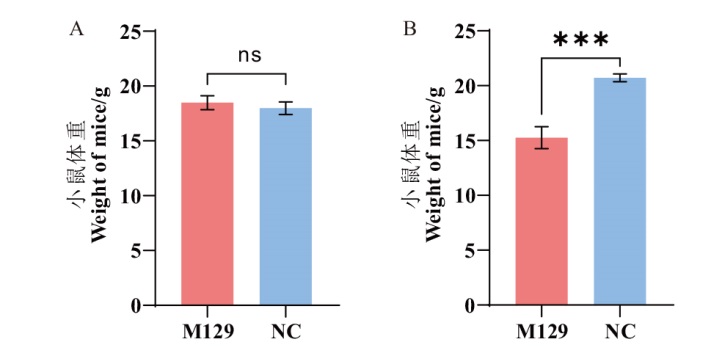
Fig. 1 Changes of mice's weight A: Weight of mice on day 0. B: Weight of mice on day 14. ***, P < 0.001; ns, nonsignificant difference. The same below
| 组别 Group | 编号 Numbering | D0体重 D0 Weight/g | D14体重 D14 Weight/g | 体重变化 Weight change/g |
|---|---|---|---|---|
| M129 | B59 | 19.0 | 15.4 | -3.6 |
| B60 | 18.5 | 15.8 | -2.7 | |
| B70 | 18.0 | 14.7 | -3.3 | |
| B72 | 17.7 | 13.9 | -3.8 | |
| B73 | 19.2 | 16.5 | -2.7 | |
| NC | B56 | 18.8 | 20.4 | 1.6 |
| B57 | 17.2 | 20.5 | 3.3 | |
| B58 | 17.8 | 21.3 | 3.5 | |
| B69 | 18.1 | 20.8 | 2.7 | |
| B71 | 18.0 | 20.6 | 2.6 |
Table 3 Record table of weights in mice
| 组别 Group | 编号 Numbering | D0体重 D0 Weight/g | D14体重 D14 Weight/g | 体重变化 Weight change/g |
|---|---|---|---|---|
| M129 | B59 | 19.0 | 15.4 | -3.6 |
| B60 | 18.5 | 15.8 | -2.7 | |
| B70 | 18.0 | 14.7 | -3.3 | |
| B72 | 17.7 | 13.9 | -3.8 | |
| B73 | 19.2 | 16.5 | -2.7 | |
| NC | B56 | 18.8 | 20.4 | 1.6 |
| B57 | 17.2 | 20.5 | 3.3 | |
| B58 | 17.8 | 21.3 | 3.5 | |
| B69 | 18.1 | 20.8 | 2.7 | |
| B71 | 18.0 | 20.6 | 2.6 |
| 组别Group | 背侧面Dorsal surface | 腹侧面Ventral surface | 编号Numbering | 评分Score | 均分Average score |
|---|---|---|---|---|---|
| M129 | 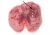 | 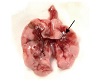 | B59 | 1 | 1.6 |
| B60 | 1 | ||||
| B70 | 2 | ||||
| B72 | 3 | ||||
| B73 | 1 | ||||
| NC | 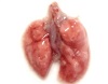 |  | B56 | 0 | 0 |
| B57 | 0 | ||||
| B58 | 0 | ||||
| B69 | 0 | ||||
| B71 | 0 |
Table 4 Gross pathological changes and scoring table of mice's lungs
| 组别Group | 背侧面Dorsal surface | 腹侧面Ventral surface | 编号Numbering | 评分Score | 均分Average score |
|---|---|---|---|---|---|
| M129 | | | B59 | 1 | 1.6 |
| B60 | 1 | ||||
| B70 | 2 | ||||
| B72 | 3 | ||||
| B73 | 1 | ||||
| NC | | | B56 | 0 | 0 |
| B57 | 0 | ||||
| B58 | 0 | ||||
| B69 | 0 | ||||
| B71 | 0 |
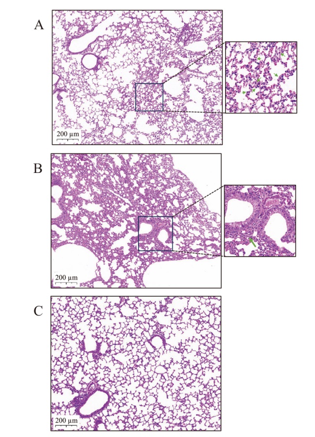
Fig. 2 Tissue sections of mice's lungs A, B: HE staining(100×)in the lungs of mice in the group M129, Figure A showed neutrophil infiltration and alveolar septum thickening, and Figure B showed inflammatory cell infiltration around the trachea(green arrow). C: HE staining(100×)in the lungs of mice in the group NC showed complete alveolar and tracheal structures and no inflammatory cell infiltration
| 组别 Group | 编号 Numbering | 各指标得分Scores for each indicator | 总分 Total score | ||||
|---|---|---|---|---|---|---|---|
| A | B | C | D | E | |||
| M129 | B59 | 0 | 2 | 0 | 0 | 0.6 | 0.292 |
| B60 | 0 | 2 | 0 | 0 | 0.6 | 0.292 | |
| B70 | 0 | 2 | 0 | 0 | 0.7 | 0.294 | |
| B72 | 0 | 2 | 0 | 0 | 0.7 | 0.294 | |
| B73 | 0 | 2 | 0 | 0 | 0.6 | 0.292 | |
| NC | B56 | 0 | 0.8 | 0 | 0 | 0 | 0.112 |
| B57 | 0 | 0.6 | 0 | 0 | 0 | 0.084 | |
| B58 | 0 | 0.7 | 0 | 0 | 0 | 0.098 | |
| B69 | 0 | 0.6 | 0 | 0 | 0 | 0.084 | |
| B71 | 0 | 0.3 | 0 | 0 | 0 | 0.042 | |
Table 5 Histopathological score results of ATS lung injury
| 组别 Group | 编号 Numbering | 各指标得分Scores for each indicator | 总分 Total score | ||||
|---|---|---|---|---|---|---|---|
| A | B | C | D | E | |||
| M129 | B59 | 0 | 2 | 0 | 0 | 0.6 | 0.292 |
| B60 | 0 | 2 | 0 | 0 | 0.6 | 0.292 | |
| B70 | 0 | 2 | 0 | 0 | 0.7 | 0.294 | |
| B72 | 0 | 2 | 0 | 0 | 0.7 | 0.294 | |
| B73 | 0 | 2 | 0 | 0 | 0.6 | 0.292 | |
| NC | B56 | 0 | 0.8 | 0 | 0 | 0 | 0.112 |
| B57 | 0 | 0.6 | 0 | 0 | 0 | 0.084 | |
| B58 | 0 | 0.7 | 0 | 0 | 0 | 0.098 | |
| B69 | 0 | 0.6 | 0 | 0 | 0 | 0.084 | |
| B71 | 0 | 0.3 | 0 | 0 | 0 | 0.042 | |
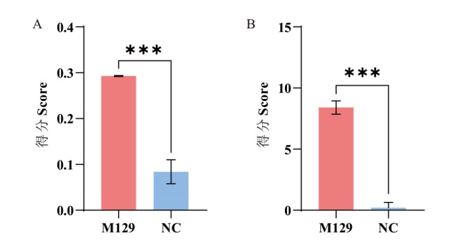
Fig. 3 Scoring results of histopathology A: Histopathological score results of ATS lung injury. B: Histopathological score results of 26-score method
| 组别 Group | 编号 Numbering | 各指标得分Scores for each indicator | 总分 Total Score | ||||
|---|---|---|---|---|---|---|---|
| A | B | C | D | E | |||
| M129 | B59 | 1 | 1 | 0 | 2 | 3 | 9 |
| B60 | 1 | 1 | 0 | 1 | 3 | 8 | |
| B70 | 1 | 1 | 0 | 1 | 3 | 8 | |
| B72 | 1 | 1 | 0 | 2 | 3 | 9 | |
| B73 | 1 | 1 | 0 | 1 | 3 | 8 | |
| NC | B56 | 1 | 0 | 0 | 0 | 0 | 1 |
| B57 | 0 | 0 | 0 | 0 | 0 | 0 | |
| B58 | 0 | 0 | 0 | 0 | 0 | 0 | |
| B69 | 0 | 0 | 0 | 0 | 0 | 0 | |
| B71 | 0 | 0 | 0 | 0 | 0 | 0 | |
Table 6 Scoring results of histopathology by 26-score method
| 组别 Group | 编号 Numbering | 各指标得分Scores for each indicator | 总分 Total Score | ||||
|---|---|---|---|---|---|---|---|
| A | B | C | D | E | |||
| M129 | B59 | 1 | 1 | 0 | 2 | 3 | 9 |
| B60 | 1 | 1 | 0 | 1 | 3 | 8 | |
| B70 | 1 | 1 | 0 | 1 | 3 | 8 | |
| B72 | 1 | 1 | 0 | 2 | 3 | 9 | |
| B73 | 1 | 1 | 0 | 1 | 3 | 8 | |
| NC | B56 | 1 | 0 | 0 | 0 | 0 | 1 |
| B57 | 0 | 0 | 0 | 0 | 0 | 0 | |
| B58 | 0 | 0 | 0 | 0 | 0 | 0 | |
| B69 | 0 | 0 | 0 | 0 | 0 | 0 | |
| B71 | 0 | 0 | 0 | 0 | 0 | 0 | |
| 组别 Group | 编号 Numbering | 平均Ct值 Average Ct value | 病原载量 Pathogen load/(CFU·mL-1) |
|---|---|---|---|
| M129 | B59 | - | - |
| B60 | - | - | |
| B70 | - | - | |
| B72 | 33.91 | 6.25 | |
| B73 | |||
| NC | B56 | - | - |
| B57 | 36.5 | 1.09 | |
| B58 | - | - | |
| B69 | - | - | |
| B71 | - | - |
Table 7 Detection of Ct value and corresponding M. pneu-moniae pathogen load in each sample
| 组别 Group | 编号 Numbering | 平均Ct值 Average Ct value | 病原载量 Pathogen load/(CFU·mL-1) |
|---|---|---|---|
| M129 | B59 | - | - |
| B60 | - | - | |
| B70 | - | - | |
| B72 | 33.91 | 6.25 | |
| B73 | |||
| NC | B56 | - | - |
| B57 | 36.5 | 1.09 | |
| B58 | - | - | |
| B69 | - | - | |
| B71 | - | - |
| 组别Group | 细胞分群情况Cell distribution situation | 编号Numbering | CD4+/CD3+ | CD8+/CD3+ | CD4+/CD8+ |
|---|---|---|---|---|---|
| M129 | 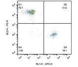 | B59 | 0.799 | 0.194 | 4.12 |
| B60 | 0.797 | 0.192 | 4.15 | ||
| B70 | 0.810 | 0.175 | 4.63 | ||
| B72 | 0.814 | 0.176 | 4.63 | ||
| B73 | 0.797 | 0.191 | 4.17 | ||
| NC | 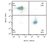 | B56 | 0.768 | 0.222 | 3.46 |
| B57 | 0.738 | 0.255 | 2.89 | ||
| B58 | 0.753 | 0.237 | 3.18 | ||
| B69 | 0.758 | 0.236 | 3.21 | ||
| B71 | 0.745 | 0.244 | 3.05 |
Table 8 Distribution and proportion of CD4 and CD8 cells in peripheral blood of mice
| 组别Group | 细胞分群情况Cell distribution situation | 编号Numbering | CD4+/CD3+ | CD8+/CD3+ | CD4+/CD8+ |
|---|---|---|---|---|---|
| M129 | | B59 | 0.799 | 0.194 | 4.12 |
| B60 | 0.797 | 0.192 | 4.15 | ||
| B70 | 0.810 | 0.175 | 4.63 | ||
| B72 | 0.814 | 0.176 | 4.63 | ||
| B73 | 0.797 | 0.191 | 4.17 | ||
| NC | | B56 | 0.768 | 0.222 | 3.46 |
| B57 | 0.738 | 0.255 | 2.89 | ||
| B58 | 0.753 | 0.237 | 3.18 | ||
| B69 | 0.758 | 0.236 | 3.21 | ||
| B71 | 0.745 | 0.244 | 3.05 |
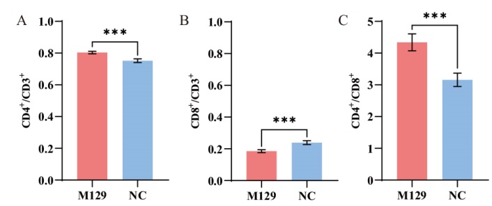
Fig. 4 Changes in the ratio of CD4+/CD3+, CD8+/CD3+, and CD4+/CD8+
| 组别Group | 编号Numbering | IL-6/(pg·mL-1) |
|---|---|---|
| M129 | B59 | 123.35 |
| B60 | 96.36 | |
| B70 | 78.35 | |
| B72 | 86.65 | |
| B73 | 46.19 | |
| NC | B56 | 10.59 |
| B57 | 16.08 | |
| B58 | 11.68 | |
| B69 | 17.43 | |
| B71 | 23.29 |
Table 9 Expressions of IL-6 in the serum of mouse
| 组别Group | 编号Numbering | IL-6/(pg·mL-1) |
|---|---|---|
| M129 | B59 | 123.35 |
| B60 | 96.36 | |
| B70 | 78.35 | |
| B72 | 86.65 | |
| B73 | 46.19 | |
| NC | B56 | 10.59 |
| B57 | 16.08 | |
| B58 | 11.68 | |
| B69 | 17.43 | |
| B71 | 23.29 |
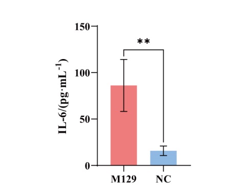
Fig. 5 Expressions of IL-6 in the serum of mouse **, P < 0.01
| [1] | Yang MC, Su YT, Chen PH, et al. Changing patterns of infectious diseases in children during the COVID-19 pandemic[J]. Front Cell Infect Microbiol, 2023, 13: 1200617. |
| [2] |
Li T, Chu C, Wei BY, et al. Immunity debt: hospitals need to be prepared in advance for multiple respiratory diseases that tend to co-occur[J]. Biosci Trends, 2024, 17(6): 499-502.
doi: 10.5582/bst.2023.01303 pmid: 38072445 |
| [3] |
Meyer Sauteur PM, Beeton ML, ESGMAC the ESGMAC MAPS study group. Mycoplasma pneumoniae: gone forever?[J]. Lancet Microbe, 2023, 4(10): e763.
doi: 10.1016/S2666-5247(23)00182-9 pmid: 37393927 |
| [4] | Yan C, Xue GH, Zhao HQ, et al. Current status of Mycoplasma pneumoniae infection in China[J]. World J Pediatr, 2024, 20(1): 1-4. |
| [5] | Li H, Li SK, Yang HJ, et al. Resurgence of Mycoplasma pneumonia by macrolide-resistant epidemic clones in China[J]. Lancet Microbe, 2024, 5(6): e515. |
| [6] | 吴移谋, 邵国青. 支原体学[M]. 3版. 北京: 人民卫生出版社, 2022. |
| Wu YM, Shao GQ. Mycoplasma[M]. 3rd ed. Beijing: People's Medical Publishing House, 2022. | |
| [7] | 王春莉, 孙锦涵, 黑志平, 等. 989例儿童肺炎支原体感染的临床特征分析[J]. 宁夏医科大学学报, 2023, 45(6): 612-618. |
| Wang CL, Sun JH, Hei ZP, et al. Clinical characteristics of 989 cases of Mycoplasma pneumoniae in children with lower respiratory tract infections[J]. J Ningxia Med Univ, 2023, 45(6): 612-618. | |
| [8] | 伊丽萍, 薛建, 任少龙, 等. 儿童肺炎支原体感染的临床特征及混合感染相关因素研究[J]. 中华流行病学杂志, 2022, 43(9): 1448-1454. |
|
Yi LP, Xue J, Ren SL, et al. Clinical characteristics of Mycoplasma pneumoniae infection and factors associated with co-infections in children[J]. Chin J Epidemiol, 2022, 43(9): 1448-1454.
doi: 10.3760/cma.j.cn112338-20220321-00210 pmid: 36117353 |
|
| [9] |
胡海洋, 应婉琴, 何军, 等. 酶促重组等温扩增实时荧光法快速检测肺炎支原体方法的建立及应用[J]. 生物技术通报, 2022, 38(9): 264-270.
doi: 10.13560/j.cnki.biotech.bull.1985.2021-1529 |
| Hu HY, Ying WQ, He J, et al. Establishment and application of ERA real-time fluorescence method for rapid detection of Mycoplasma pneumoniae[J]. Biotechnol Bull, 2022, 38(9): 264-270. | |
| [10] | Chen YC, Hsu WY, Chang TH. Macrolide-resistant Mycoplasma pneumoniae infections in pediatric community-acquired pneumonia[J]. Emerg Infect Dis, 2020, 26(7): 1382-1391. |
| [11] | Tsai TA, Tsai CK, Kuo KC, et al. Rational stepwise approach for Mycoplasma pneumoniae pneumonia in children[J]. J Microbiol Immunol Infect, 2021, 54(4): 557-565. |
| [12] | Jiang ZL, Li SH, Zhu CM, et al. Mycoplasma pneumoniae infections: pathogenesis and vaccine development[J]. Pathogens, 2021, 10(2): 119. |
| [13] | Maselli DJ, Medina JL, Brooks EG, et al. The immunopathologic effects of Mycoplasma pneumoniae and community-acquired respiratory distress syndrome toxin. A primate model[J]. Am J Respir Cell Mol Biol, 2018, 58(2): 253-260. |
| [14] | Hayakawa M, Taguchi H, Kamiya S, et al. Animal model of My-coplasma pneumoniae infection using germfree mice[J]. Clin Diagn Lab Immunol, 2002, 9(3): 669-676. |
| [15] | Fonseca-Aten M, Ríos AM, Mejías A, et al. Mycoplasma pneumoniae induces host-dependent pulmonary inflammation and airway obstruction in mice[J]. Am J Respir Cell Mol Biol, 2005, 32(3): 201-210. |
| [16] | Tamiya S, Yoshikawa E, Suzuki K, et al. Susceptibility analysis in several mouse strains reveals robust T-cell responses after My-coplasma pneumoniae infection in DBA/2 mice[J]. Front Cell Infect Microbiol, 2021, 10: 602453. |
| [17] | Wu YZ, Yu YF, Hua LZ, et al. Genotyping and biofilm formation of Mycoplasma hyopneumoniae and their association with virulence[J]. Vet Res, 2022, 53(1): 95. |
| [18] | Matute-Bello G, Downey G, Moore BB, et al. An official American Thoracic Society workshop report: features and measurements of experimental acute lung injury in animals[J]. Am J Respir Cell Mol Biol, 2011, 44(5): 725-738. |
| [19] |
Cimolai N, Taylor GP, Mah D, et al. Definition and application of a histopathological scoring scheme for an animal model of acute Mycoplasma pneumoniae pulmonary infection[J]. Microbiol Immunol, 1992, 36(5): 465-478.
pmid: 1513263 |
| [20] | 孟凡亮, 何利华, 顾一心, 等. 实时荧光定量-聚合酶链反应方法检测肺炎支原体[J]. 疾病监测, 2013, 28(3): 209-212. |
| Meng FL, He LH, Gu YX, et al. A real-time PCR assay for detection of Mycoplasma pneumoniae[J]. Dis Surveillance, 2013, 28(3): 209-212. | |
| [21] | 章曼曼, 林立, 李昌崇. 儿童社区获得性肺炎病原及混合感染研究进展[J]. 中国实用儿科杂志, 2019, 34(12): 1034-1037. |
| Zhang MM, Lin L, Li CC. Research progress in the etiology of community-acquired pneumonia and coinfection in children[J]. Chin J Pract Pediatr, 2019, 34(12): 1034-1037. | |
| [22] | Atkinson TP, Balish MF, Waites KB. Epidemiology, clinical manifestations, pathogenesis and laboratory detection of Mycoplasma pneumoniae infections[J]. FEMS Microbiol Rev, 2008, 32(6): 956-973. |
| [23] | Hu J, Ye YY, Chen XX, et al. Insight into the pathogenic mechanism of Mycoplasma pneumoniae[J]. Curr Microbiol, 2022, 80(1): 14. |
| [24] | Techasaensiri C, Tagliabue C, Cagle M, et al. Variation in colonization, ADP-ribosylating and vacuolating cytotoxin, and pulmonary disease severity among Mycoplasma pneumoniae strains[J]. Am J Respir Crit Care Med, 2010, 182(6): 797-804. |
| [25] | 吴振起, 杨璐, 敏娜, 等. 清燥救肺汤及其拆方对肺炎支原体感染小鼠Bax、Bcl-2、Caspase-3蛋白的影响[J]. 中草药, 2018, 49(2): 389-395. |
| Wu ZQ, Yang L, Min N, et al. Effect of Qingzao Jiufei Decoction and its decomposing agent on MP infection Bax, Bcl-2, and Caspase-3[J]. Chin Tradit Herb Drugs, 2018, 49(2): 389-395. | |
| [26] | Sibila M, Pieters M, Molitor T, et al. Current perspectives on the diagnosis and epidemiology of Mycoplasma hyopneumoniae infection[J]. Vet J, 2009, 181(3): 221-231. |
| [27] | Kang SJ, Narazaki M, Metwally H, et al. Historical overview of the interleukin-6 family cytokine[J]. J Exp Med, 2020, 217(5): e20190347. |
| [28] | Tanaka T, Narazaki M, Kishimoto T. IL-6 in inflammation, immunity, and disease[J]. Cold Spring Harb Perspect Biol, 2014, 6(10): a016295. |
| [29] | Yang J, Hooper WC, Phillips DJ, et al. Cytokines in Mycoplasma pneumoniae infections[J]. Cytokine Growth Factor Rev, 2004, 15(2/3): 157-168. |
| [30] |
宗阳, 姚卫峰, 居文政. 以白介素6为受体挖掘中药单体治疗新型冠状病毒肺炎引发的细胞因子风暴的干预作用[J]. 中国医院药学杂志, 2020, 40(11): 1182-1188.
doi: 10.13286/j.1001-5213.2020.11.02 |
|
Zong Y, Yao WF, Ju WZ. The intervention effect investigation of Chinese medicine monomer on cytokine storm induced by COVID-19 based on interleukin-6 receptor[J]. Chin J Hosp Pharm, 2020, 40(11): 1182-1188.
doi: 10.13286/j.1001-5213.2020.11.02 |
|
| [31] | Forcina L, Franceschi C, Musarò A. The hormetic and hermetic role of IL-6[J]. Ageing Res Rev, 2022, 80: 101697. |
| [32] | Romero-Rojas A, Ponce-Hernández C, Mendoza SE, et al. Immunomodulatory properties of Mycoplasma pulmonis[J]. Int Immunopharmacol, 2001, 1(9/10): 1689-1697. |
| [33] | 沈小芳. 支炎合剂对肺炎支原体肺炎模型大鼠CD4+T、CD8+ T细胞的影响[D]. 长沙: 湖南中医药大学, 2021. |
| Shen XF. Effect of zhiyan mixture on CD4+ T and CD8+ T cells in rats with Mycoplasma pneumonia[D]. Changsha: Hunan University of Chinese Medicine, 2021. | |
| [34] | Dumke R, Catrein I, Herrmann R, et al. Preference, adaptation and survival of Mycoplasma pneumoniae subtypes in an animal model[J]. Int J Med Microbiol, 2004, 294(2/3): 149-155. |
| [35] | Lai CC, Hsueh CC, Hsu CK, et al. Disease burden and macrolide resistance of Mycoplasma pneumoniae infection in adults in the Asia-Pacific region[J]. Int J Antimicrob Agents, 2024, 64(2): 107205. |
| [36] | Xu X, Greenland J, Baluk P, et al. Cathepsin L protects mice from mycoplasmal infection and is essential for airway lymphangiogenesis[J]. Am J Respir Cell Mol Biol, 2013, 49(3): 437-444. |
| [37] | Martin RJ, Chu HW, Honour JM, et al. Airway inflammation and bronchial hyperresponsiveness after Mycoplasma pneumoniae infection in a murine model[J]. Am J Respir Cell Mol Biol, 2001, 24(5): 577-582. |
| [38] |
Boonyarattanasoonthorn T, Elewa YHA, Tag-El-Din-Hassan HT, et al. Profiling of cellular immune responses to Mycoplasma pulmonis infection in C57BL/6 and DBA/2 mice[J]. Infect Genet Evol, 2019, 73: 55-65.
doi: S1567-1348(19)30058-9 pmid: 31026602 |
| [1] | YU Yong-xia, ZHU Ning, LIU Guang-min, ZHU Long-jiao, XU Wen-tao. Research Progress in Nucleic Acid Molecular Diagnostic Technology for Mycoplasma pneumoniae [J]. Biotechnology Bulletin, 2024, 40(12): 72-83. |
| [2] | HU Hai-yang, YING Wan-qin, HE Jun, LV Zhi-xian, XIE Xiao-ping, DENG Zhong-liang. Establishment and Application of ERA Real-time Fluorescence Method for Rapid Detection of Mycoplasma pneumoniae [J]. Biotechnology Bulletin, 2022, 38(9): 264-270. |
| Viewed | ||||||
|
Full text |
|
|||||
|
Abstract |
|
|||||