生物技术通报 ›› 2021, Vol. 37 ›› Issue (10): 137-142.doi: 10.13560/j.cnki.biotech.bull.1985.2020-1509

• 研究报告 • 上一篇    下一篇

牛病毒性腹泻病毒离子孔道蛋白p7多肽多克隆抗体的制备和鉴定

付强(), 郭妍婷, 陈俊贞, 王金泉, 史慧君()   

  1. 新疆农业大学动物医学学院,乌鲁木齐 830052
  • 收稿日期:2020-12-13 出版日期:2021-10-26 发布日期:2021-11-12
  • 作者简介:付强,男,副教授,研究方向:病原微生物致病机制;E-mail: 466183013@qq.com;
  • 基金资助:
    国家自然科学基金项目(31902271);国家自然科学基金项目(32060042);自治区天山创新团队(2020D14005);新疆农业大学研究生创新项目(XJAUGRI2020023)

Preparation and Identification of Polyclonal Antibody Against Ion Channel Protein p7 Polypeptide of Bovine Viral Diarrhea Virus

FU Qiang(), GUO Yan-ting, CHEN Jun-zhen, WANG Jin-quan, SHI Hui-jun()   

  1. College of Veterinary Medicine,Xinjiang Agricultural University,Urumqi 830052
  • Received:2020-12-13 Published:2021-10-26 Online:2021-11-12

摘要:

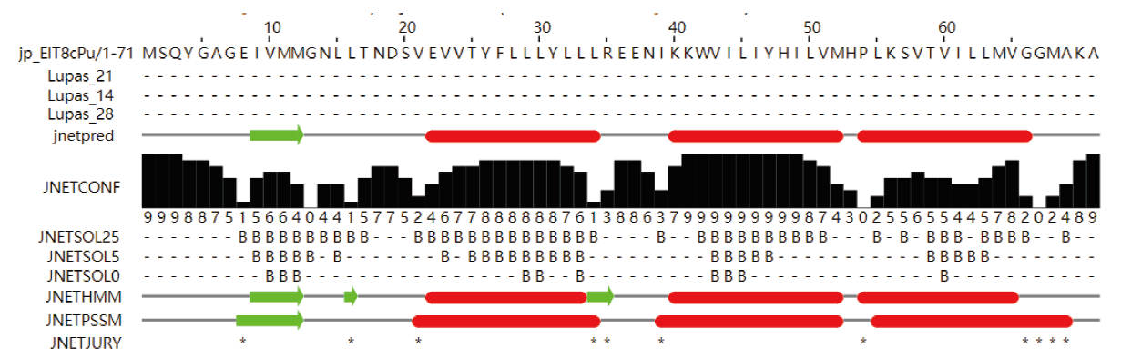
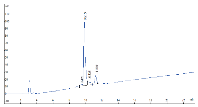
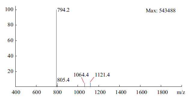
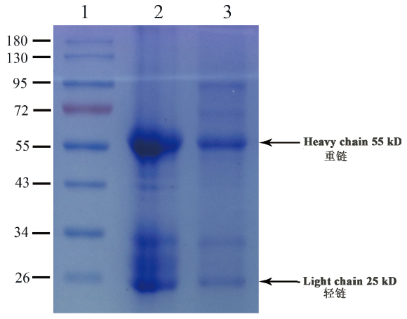
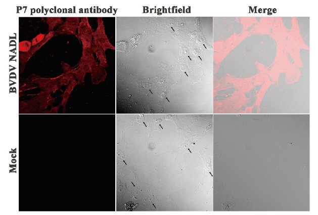
牛病毒性腹泻病毒(Bovine viral diarrhea virus,BVDV)非结构蛋白p7因其离子通道活性被称为病毒孔蛋白,参与病毒进入、释放和复制的过程。为了深入研究p7蛋白形成离子通道的机理,使用人工合成的多肽制备p7特异性多克隆抗体。根据GenBank中BVDV毒株NADL p7氨基酸序列信息,进行生物信息学分析,合成表面抗原多肽并偶联血蓝蛋白(keyhole limpet hemocyanin,KLH)作为载体蛋白,经反相高效液相色谱(reversed-phase-high performance liquid chromatography,RP-HPLC)纯化、纯度分析,用质谱进行定性鉴定;使用弗氏佐剂进行乳化后,对新西兰大白兔进行皮下多点注射免疫,免疫剂量为400 μg/只,连续5次免疫,免疫后10 d时采用多肽亲和柱纯化血清,获得多肽多克隆抗体,使用间接ELISA和SDS-PAGE进行抗体效价和纯度检测;BVDV NADL感染牛肾细胞MDBK后使用免疫荧光染色检测多肽多克隆抗体的反应性和特异性。生物信息学分析p7蛋白的主要抗原表位分布在氨基端,偶联KLH后合成多肽MSQYGAGEIVM MGN-Cys-KLH,经RP-HPLC纯化后多肽纯度可达80.19%,分子量为794.2 Da;经过5次免疫并使用多肽亲和柱纯化血清后,间接ELISA检测多肽多克隆抗体效价为1:100 000,Western blot检测抗体的纯度为90.2%;使用免疫荧光染色检测发现该抗体在BVDV NADL感染的MDBK细胞的细胞膜上呈现阳性染色,与p7离子孔道定位相符。成功制备兔抗BVDV p7多肽多克隆抗体,具有较好的反应性和特异性,为进一步研究p7形成离子孔道的机理提供了有利工具。

关键词: 牛病毒性腹泻病毒, p7蛋白, 离子孔道蛋白, 多肽, 多克隆抗体

Abstract:

The non-structural protein p7 of bovine viral diarrhea virus(BVDV)is called viroporin due to its ion channel activity,which participates in the process of virus entry,release and replication. To further investigate the mechanism of p7 protein forming ion channel,p7 specific polyclonal antibody was prepared using the synthetic peptide. According to the amino acid information of NADL p7 of BVDV strain in GenBank,the bioinformatics analysis was conducted,and the surface antigen polypeptide was synthesized and conjugated with the protein of keyhole limpet hemocyanin(KLH)served as the carrier protein. The protein was purified by reversed-phase-high performance liquid chromatography(RP-HPLC),the purity was analysed,and qualitative identification was conducted by mass spectrometry. Then the peptide plus Freund’s adjuvant was emulsificated and multiple subcutaneously injected into the New Zealand white rabbit,and the immune dose was 400 μg per rabbit,which repeated continuously 5 times. The serum was purified by peptide affinity column at 10 d after immunization,and the polypeptide polyclonal antibody was acquired. Indirect ELISA and SDS-PAGE were applied to detect the titer and purity of the polyclonal antibody. Immunofluorescence staining was used to detect the reactivity and specificity of the polyclonal antibody after BVDV NADL infected the MDBK of bovine renal cells. Bioinformatics analysis showed that the main antigenic epitopes were located in the N-terminal of p7 protein. The peptide MSQYGAGEIVMMGN-Cys-KLH was synthesized after KLH conjugation. The purity of the peptide reached 80.19% and the molecular weight was 794.2 Da. After 5 times of immunization and the serum was purified using the peptide affinity column,the titer of the polyclonal antibody detected by indirect ELISA was 1:100 000,and the antibody purity detected by Western blot was 90.2%. Immunofluorescence staining showed that the staining of the polyclonal antibody was positive on the membrane of MDBK cells infected with BVDV NADL,which was consistent with the location of p7 ion channel. The rabbit anti-BVDV p7 polypeptide polyclonal antibody was successfully prepared,and had high reactivity and specificity,which provides a useful tool for further investigation on the mechanism of p7 forming ion channels.

Key words: bovine viral diarrhea virus, p7 protein, ion channel protein, polypeptide, polyclonal antibody