


Biotechnology Bulletin ›› 2022, Vol. 38 ›› Issue (6): 279-290.doi: 10.13560/j.cnki.biotech.bull.1985.2021-1152
Previous Articles Next Articles
WANG Nan( ), ZHANG Rui, PAN Yang-yang, HE Hong-hong, WANG Jing-lei, CUI Yan, YU Si-jiu()
Received:2021-09-07
Online:2022-06-26
Published:2022-07-11
Contact:
YU Si-jiu
E-mail:594302635@qq.com;sjyu@163.com
WANG Nan, ZHANG Rui, PAN Yang-yang, HE Hong-hong, WANG Jing-lei, CUI Yan, YU Si-jiu. Cloning of Bos grunniens TGF-β1 Gene and Its Expression in Major Organs of Female Reproductive System[J]. Biotechnology Bulletin, 2022, 38(6): 279-290.
| 引物Primer | 引物序列Primer sequence(5'-3') | 产物长度Product size/bp | 退火温度Annealing temperature Tm/℃ | GenBank登录号GenBank No. |
|---|---|---|---|---|
| D-TGF-β1 | F:ACTACTACGCCAAGGAGGTCAC | 153 | 60 | MZ004937 |
| R:TTCCGAGAATCTTAGCTGCA | ||||
| TGF-β1 | F:GCAAACAGACCCTCCTACCTTT | 1 173 | 60 | MZ004937 |
| R:TCCTTAAATACAGTCCTGGTGAG | ||||
| GAPDH | F:GGGTCATCATCTCTGCACCT | 178 | 60 | EU195062.1 |
| R:TGGTCATAAGTCCCTCCACG |
Table 1 Primer sequences and length
| 引物Primer | 引物序列Primer sequence(5'-3') | 产物长度Product size/bp | 退火温度Annealing temperature Tm/℃ | GenBank登录号GenBank No. |
|---|---|---|---|---|
| D-TGF-β1 | F:ACTACTACGCCAAGGAGGTCAC | 153 | 60 | MZ004937 |
| R:TTCCGAGAATCTTAGCTGCA | ||||
| TGF-β1 | F:GCAAACAGACCCTCCTACCTTT | 1 173 | 60 | MZ004937 |
| R:TCCTTAAATACAGTCCTGGTGAG | ||||
| GAPDH | F:GGGTCATCATCTCTGCACCT | 178 | 60 | EU195062.1 |
| R:TGGTCATAAGTCCCTCCACG |
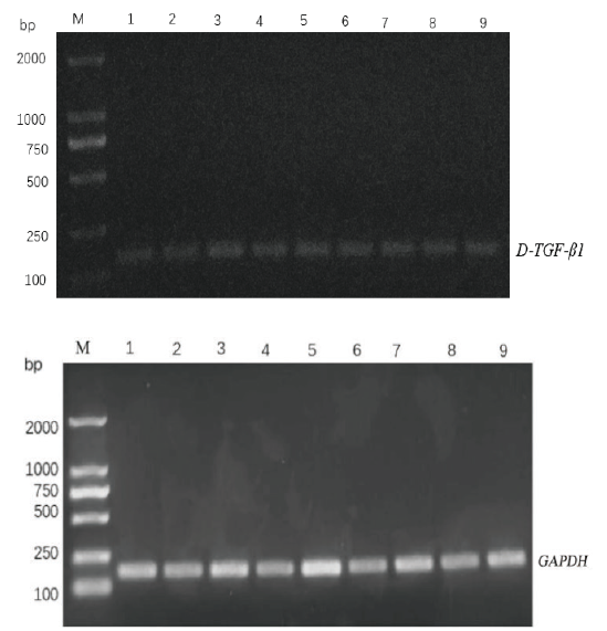
Fig. 1 D-TGF-β1 and GAPDH PCR amplification electrop-horesis M:DL2000 DNA marker. 1-3:Fallopian tubes,uterus and ovaries in luteal phase. 4-6:Fallopian tubes,uterus and ovaries in pregnancy phase. 7-8:Fallopian tubes,uterus and ovaries in follicular phase
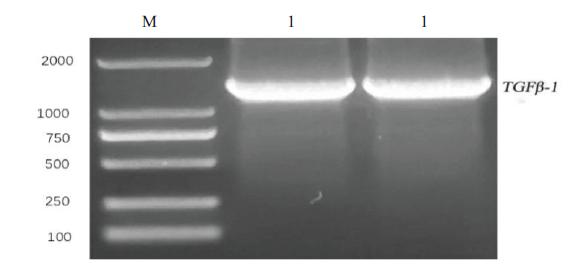
Fig. 2 TGF-β1 PCR amplification electrophoresis M:DL2000 DNA marker. 1:Fallopian tubes

Fig. 3 Sequence alignment of TGF-β1 gene among different species Black arrow points to nucleotide 414 and 1 168
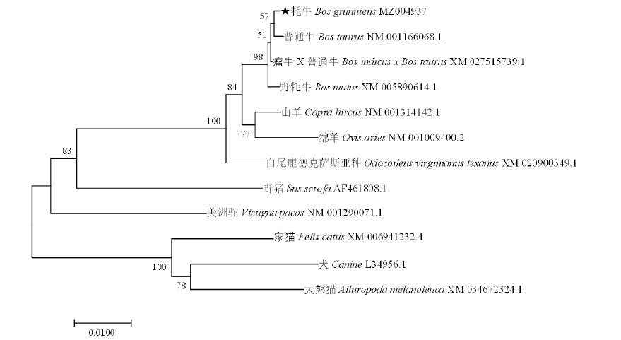
Fig.4 Phylogenetic tree of TGF-β1gene “★”indicates that the data is obtained from this experiment
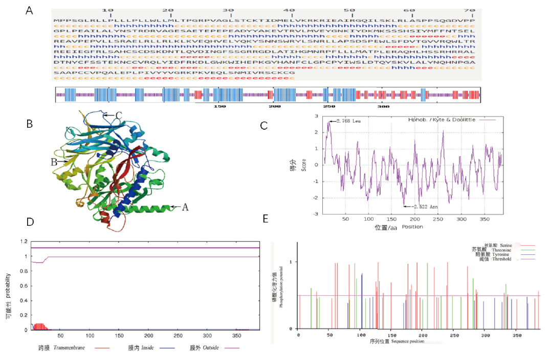
Fig. 5 Results of yak TGF-β1 protein via bioinformatics analysis A:The secondary structure prediction of TGF-β1 protein in Bos grunniens. The long vertical zone:Alpha helix(h). The middle vertical zone:Extended stand(e). The short vertical zone:Random coil(c). B:The tertiary structure prediction of TGF-β1 protein in Bos grunniens. C:Hydrophilicity and hydrophobicity of the protein encoded by TGF-β1 gene. D:The protein transmembrane regional analysis of TGF-β1 protein in Bos grunniens. E:TGF-β1 phosphorylation site analyses
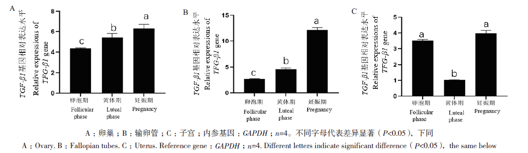
Fig. 6 Relative expressions of TGF-β1 mRNA in different tissues A:Ovary. B:Fallopian tubes. C:Uterus. Reference gene:GAPDH;n=4. Different letters indicate significant difference(P<0.05),the same below
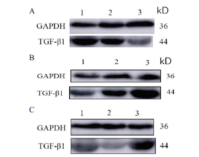
Fig. 7 Detection results of TGF-β1 and GAPDH protein in different tissues A:Ovary. B:Fallopian tubes. C:Uterus. 1:Follicular phase. 2:Luteal phase. 3:Pregnancy
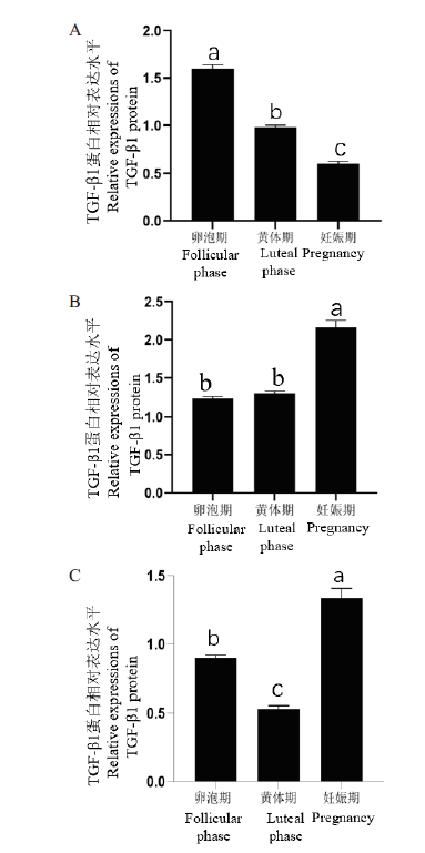
Fig. 8 Relative expressions of TGF-β1 protein in different tissues A:Ovary. B:Fallopian tubes. C:Uterus. Reference protein:GAPDH;n=4

Fig. 9 Distribution of TGF-β1 proteins in different tissues A-D:Positive expression. A,A1,A2,A3,B,B1,and B2:Ovary. C,C1,and C2:Fallopian tube. D,D1,D2:Uterus. E-H:Negative control.SG:Follicle granular layer. EM:Mucosalepithelium. GE:Germinal epitnelium. TF:Theca follicle. CL:Corpus luteum verum. EP:Epitheliummucosae.UG:Uterineglands. sg:Serous gland
| [1] | 汪琦. 牦牛四个低氧适应基因的遗传多态性研究[D]. 成都: 西南民族大学, 2017. |
| Wang Q. Study on genetic polymorphisms of four hypoxia adaptation genes in yak[D]. Chengdu: Southwest University for Nationalities, 2017. | |
| [2] |
Qiu Q, Zhang GJ, Ma T, et al. The yak genome and adaptation to life at high altitude[J]. Nat Genet, 2012, 44(8):946-949.
doi: 10.1038/ng.2343 URL |
| [3] |
Rowe P. Clinical potential for TGF-Β[J]. Lancet, 1994, 344(8915):72-73.
pmid: 7912386 |
| [4] |
Miyazono K, Kusanagi K, Inoue H. Divergence and convergence of TGF-? /BMP signaling[J]. J Cell Physiol, 2001, 187(3):265-276.
doi: 10.1002/jcp.1080 pmid: 11319750 |
| [5] |
Moustakas A, Pardali K, Gaal A, et al. Mechanisms of TGF-beta signaling in regulation of cell growth and differentiation[J]. Immunol Lett, 2002, 82(1/2):85-91.
doi: 10.1016/S0165-2478(02)00023-8 URL |
| [6] |
Attisano A, Wrana JL. Signal transduction by the TGF-beta superfamily[J]. Science, 2002, 296(5573):1646-1647.
pmid: 12040180 |
| [7] | Ziyadeh FN. Mediators of diabetic renal disease:the case for tgf-Beta as the major mediator[J]. J Am Soc Nephrol, 2004, 15(Suppl 1):S55-S57. |
| [8] |
Nolan VG, Adewoye A, Baldwin C, et al. Sickle cell leg ulcers:associations with haemolysis and SNPs in Klotho, TEK and genes of the TGF-beta/BMP pathway[J]. Br J Haematol, 2006, 133(5):570-578.
doi: 10.1111/j.1365-2141.2006.06074.x URL |
| [9] | 郭永红, 罗金燕. TGF-β超家族与smad信号转导研究进展[J]. 医学综述, 2005, 11(8):685-688. |
| Guo YH, Luo JY. Advances in TGF-β superfamily and Smad signaling research[J]. Med Recapitul, 2005, 11(8):685-688. | |
| [10] |
Okazaki R, Sakai A, Nakamura T, et al. Effects of transforming growth factor beta s and basic fibroblast growth factor on articular chondrocytes obtained from immobilised rabbit knees[J]. Ann Rheum Dis, 1996, 55(3):181-186.
pmid: 8712881 |
| [11] |
Kolambkar YM, Peister A, Soker S, et al. Chondrogenic differentiation of amniotic fluid-derived stem cells[J]. J Mol Histol, 2007, 38(5):405-413.
pmid: 17668282 |
| [12] | 周玉娟. 合浦珠母贝TGFβ信号通路相关基因的克隆及功能研究[D]. 北京: 清华大学, 2010. |
| Zhou YJ. Cloning and functional studies on genes related to TGFβ signal pathways of Pinctada fucata[D]. Beijing: Tsinghua University, 2010. | |
| [13] |
Heine U, Munoz EF, Flanders KC, et al. Role of transforming growth factor-beta in the development of the mouse embryo[J]. J Cell Biol, 1987, 105(6 pt 2):2861-2876.
pmid: 3320058 |
| [14] |
Hughes FM, Gorospe WC. Biochemical identification of apoptosis(programmed cell death)in granulosa cells:evidence for a potential mechanism underlying follicular atresia[J]. Endocrinology, 1991, 129(5):2415-2422.
pmid: 1935775 |
| [15] |
Yan ZK, Shen DF, Liao JL, et al. Hypoxia suppresses TGF-B1-induced cardiac myocyte myofibroblast transformation by inhibiting Smad2/3 and rhoa signaling pathways[J]. Cell Physiol Biochem, 2018, 45(1):250-257.
doi: 10.1159/000486771 URL |
| [16] | 徐梦思. TGF β-SMAD信号通路对猪颗粒细胞和繁殖性状的作用研究[D]. 石河子: 石河子大学, 2015. |
| Xu MS. The influence of TGF beta SMAD signaling pathway on the pig granulosa cells and reproductive traits[D]. Shihezi: Shihezi University, 2015. | |
| [17] |
Massagué J. TGF-β SIGNAL TRANSDUCTION[J]. Annu Rev Biochem, 1998, 67(1):753-791.
doi: 10.1146/annurev.biochem.67.1.753 URL |
| [18] |
Grönroos E, Kingston IJ, Ramachandran A, et al. Transforming growth factor β inhibits bone morphogenetic protein-induced transcription through novel phosphorylated Smad1/5-Smad3 complexes[J]. Mol Cell Biol, 2012, 32(14):2904-2916.
doi: 10.1128/MCB.00231-12 pmid: 22615489 |
| [19] | 张瑞, 王靖雷, 潘阳阳, 等. 牦牛CAV1基因克隆及其在雌性生殖系统主要器官中的表达定位[J]. 农业生物技术学报, 2020, 28(4):681-692. |
| Zhang R, Wang JL, Pan YY, et al. Cloning of yak(Bos grunniens)CAV1 gene and its expression in major organs of female reproductive system[J]. J Agric Biotechnol, 2020, 28(4):681-692. | |
| [20] | 伍晨, 贺全勇. TGF-β1对瘢痕疙瘩形成的影响[J]. 生命的化学, 2021, 41(4):633-641. |
| Wu C, He QY. The effect of TGF-β1 on keloid formation[J]. Chem Life, 2021, 41(4):633-641. | |
| [21] |
Sporn MB, Roberts AB. Transforming growth factor-beta. Multiple actions and potential clinical applications[J]. JAMA, 1989, 262(7):938-941.
doi: 10.1001/jama.1989.03430070086036 URL |
| [22] |
Zhou S, Zhao D, Liu SQ, et al. TGF-β1 sustains germ cell cyst reservoir via restraining follicle formation in the chicken[J]. Cell Biol Int, 2020, 44(3):861-872.
doi: 10.1002/cbin.11283 URL |
| [23] | 王馥新. TGF-β1对人卵巢颗粒细胞排卵相关基因的调控及机制研究[D]. 南京: 南京医科大学, 2020. |
| Wang FX. The role of TGF-β1 in the regulation of ovulation-related genes in humuan granulosa cells[D]. Nanjing: Nanjing Medical University, 2020. | |
| [24] | 金莉萍. 转化生长因子β与生殖功能调节研究进展[J]. 国外医学:计划生育分册, 2003, 22(1):17-20. |
| Jin LP. Advances in the study of transforming growth factor beta and regulation of reproductive function[J]. Foreign Med Sci, 2003, 22(1):17-20. | |
| [25] | 张新奇. IL-26通过调控TGF-β1/smad2信号通路介导肝星状细胞的增殖和活化而促进肝纤维化[D]. 上海: 中国人民解放军海军军医大学, 2020. |
| Zhang XQ. Interleukin-26 promotes the proliferation and activation of hepatic stellate cells to exacerbate liver fibrosis via the TGF-β1/Smad2 signaling pathway[D]. Shanghai: People’s Liberation Army Naval Medical University, 2020. | |
| [26] | 王琼. HMGB1通过NF-κB激活TGF-β1诱导特发性肺纤维化发病机制的研究[D]. 南京: 南京医科大学, 2017. |
| Wang Q. Study on the pathogenesis of HMGB1-induced idiopathic pulmonary fibrosis through NF-κB activation of TGF-β1[D]. Nanjing: Nanjing Medical University, 2017. | |
| [27] | 万云鹏. 15-LOX-1调控骨性关节炎软骨下骨中TGF-β1表达的研究[D]. 合肥: 安徽医科大学, 2020. |
| Wan YP.15-lipoxygenase-1 regulates TGF-β 1 expression in osteoarthritis subchondral bone[D]. Hefei: Anhui Medical University, 2020. | |
| [28] | 田萍. CBX7通过ITGβ3/TGFβ1/AKT信号通路在宫颈癌进展中的作用机制研究[D]. 乌鲁木齐: 新疆医科大学, 2020. |
| Tian P. Exploration of the mechanism of CBX7 in the development of cervical cancer via ITGβ3/TGFβ1/AKT signal pathway[D]. Urumqi: Xinjiang Medical University, 2020. | |
| [29] | 刘新宇, 曲陆荣. TGF-β1与超促排卵胚胎发育及妊娠结局的关系[J]. 中国医科大学学报, 2005, 34(2):162-163. |
| Liu XY, Qu LR. Relationship between transforming growth factor-β1 and embryo quality and pregnancy outcome in hyper-stimulating ovarian cycles[J]. J China Med Univ, 2005, 34(2):162-163. | |
| [30] | 王建辰, 章孝荣. 动物生殖调控[M]. 合肥: 安徽科学技术出版社, 1998. |
| Wang JC, Zhang XR. Regulation of animal reproduction[M]. Hefei: Anhui Science & Technology Publishing House, 1998. | |
| [31] |
Eppig JJ. Oocyte control of ovarian follicular development and function in mammals[J]. Reproduction, 2001, 122(6):829-838.
doi: 10.1530/rep.0.1220829 pmid: 11732978 |
| [32] | 谢江燕, 何畏, 赵俪梅, 等. 卵巢早衰患者CD4+CD25+调节性T细胞的变化及干扰素-γ、转化生长因子-β1的表达[J]. 华西医学, 2013, 28(3):377-379. |
| Xie JY, He W, Zhao LM, et al. Analysis of treg change and experssion of interferon-γ and transforming growth factor-β1 in patients with premature ovarian failure[J]. West China Med J, 2013, 28(3):377-379. | |
| [33] | 袁丽娟, 任君旭, 姬宏宇, 等. 转化生长因子-β和smad4在绝经过渡期大鼠卵巢颗粒细胞中的表达[J]. 解剖学报, 2018, 49(1):108-112. |
| Yuan LJ, Ren JX, Ji HY, et al. Expression of transforming growth factor-β and Smad4 in ovarian granulosa cells in menopausal transitional rat[J]. Acta Anat Sin, 2018, 49(1):108-112. | |
| [34] | 罗丽兰. 输卵管的解剖和功能[J]. 中国实用妇科与产科杂志, 2000, 16(4):21-22. |
| Luo LL. Anatomy and function of the fallopian tube[J]. Chin J Pract Gynecol Obstet, 2000, 16(4):21-22. | |
| [35] |
Cometti BPS, Dubey RK, Imthurn B, et al. Natural and environmental oestrogens induce TGFB1 synthesis in oviduct cells[J]. Reproduction, 2018, 155(3):233-244.
doi: 10.1530/REP-17-0425 URL |
| [36] | 王靖雷, 王萌, 潘阳阳, 等. IL-1β及其受体在雌牦牛主要生殖器官和孤雌激活胚胎中的表达定位[J]. 西北农业学报, 2018, 27(10):1395-1404. |
| Wang JL, Wang M, Pan YY, et al. Expression and localization of interleukin 1 beta and interleukin 1 receptor(type I)in main reproductive organs and parthenogenetic embryos of female yak(bosgrunniens)[J]. Acta Agric Boreali Occidentalis Sin, 2018, 27(10):1395-1404. | |
| [37] | Zhao Y, Chegini N, Flanders KC. Human fallopian tube expresses transforming growth factor(TGF beta)isoforms, TGF beta type I-III receptor messenger ribonucleic acid and protein, and contains[125I]TGF beta-binding sites[J]. J Clin Endocrinol Metab, 1994, 79(4):1177-1184. |
| [38] |
Jones RL, Stoikos C, Findlay JK, et al. TGF-β superfamily expression and actions in the endometrium and placenta[J]. Reproduction, 2006, 132(2):217-232.
doi: 10.1530/rep.1.01076 URL |
| [39] | Monsivais D, Matzuk MM, Pangas SA. The TGF-β family in the reproductive tract[J]. Cold Spring Harb Perspect Biol, 2017, 9(10):a022251. |
| [40] |
Guo F, Si CC, Zhou MJ, et al. Corrigendum. Decreased PECAM1-mediated TGF-β1 expression in the mid-secretory endometrium in women with recurrent implantation failure[J]. Hum Reprod, 2020, 35(1):253.
doi: 10.1093/humrep/dez236 URL |
| [41] |
Li J, Dong XY, Yang PW, et al. Activation of uterine Smad3 pathway is crucial for embryo implantation[J]. Curr Med Sci, 2019, 39(6):997-1002.
doi: 10.1007/s11596-019-2134-z URL |
| [42] | Hill JA, Anderson DJ. Immunological mechanisms in recurrent spontaneous abortion[J]. Arch Immunol Ther Exp(Warsz), 1990, 38(1/2):111-119. |
| [43] |
Clark DA, Flanders KC, Banwatt D, et al. Murine pregnancy decidua produces a unique immunosuppressive molecule related to transforming growth factor beta-2[J]. J Immunol, 1990, 144(8):3008-3014.
pmid: 2182711 |
| [44] | 沈霞芬. 家畜组织学与胚胎学[M]. 第4版. 北京: 中国农业出版社, 2010. |
| Shen XF. Histology and embryology of domestic animals[M]. 4th ed. Beijing: China Agricultural Press, 2010. |
| [1] | LIN Hong-yan, GUO Xiao-rui, LIU Di, LI Hui, LU Hai. Molecular Mechanism of Transcriptional Factor AtbHLH68 in Regulating Cell Wall Development by Transcriptome Analysis [J]. Biotechnology Bulletin, 2023, 39(9): 105-116. |
| [2] | YANG Zhi-xiao, HOU Qian, LIU Guo-quan, LU Zhi-gang, CAO Yi, GOU Jian-yu, WANG Yi, LIN Ying-chao. Responses of Rubisco and Rubisco Activase in Different Resistant Tobacco Strains to Brown Spot Stress [J]. Biotechnology Bulletin, 2023, 39(9): 202-212. |
| [3] | CHEN Zhong-yuan, WANG Yu-hong, DAI Wei-jun, ZHANG Yan-min, YE Qian, LIU Xu-ping, TAN Wen-Song, ZHAO Liang. Mechanism Investigation of Ferric Ammonium Citrate on Transfection for Suspended HEK293 Cells [J]. Biotechnology Bulletin, 2023, 39(9): 311-318. |
| [4] | LYU Qiu-yu, SUN Pei-yuan, RAN Bin, WANG Jia-rui, CHEN Qing-fu, LI Hong-you. Cloning, Subcellular Localization and Expression Analysis of the Transcription Factor Gene FtbHLH3 in Fagopyrum tataricum [J]. Biotechnology Bulletin, 2023, 39(8): 194-203. |
| [5] | WANG Jia-rui, SUN Pei-yuan, KE Jin, RAN Bin, LI Hong-you. Cloning and Expression Analyses of C-glycosyltransferase Gene FtUGT143 in Fagopyrum tataricum [J]. Biotechnology Bulletin, 2023, 39(8): 204-212. |
| [6] | LI Bo, LIU He-xia, CHEN Yu-ling, ZHOU Xing-wen, ZHU Yu-lin. Cloning, Subcellular Localization and Expression Analysis of CnbHLH79 Transcription Factor from Camellia nitidissima [J]. Biotechnology Bulletin, 2023, 39(8): 241-250. |
| [7] | WANG Shuai, FENG Yu-mei, BAI Miao, DU Wei-jun, YUE Ai-qin. Functional Analysis of Soybean Gene GmHMGR Responding to Exogenous Hormones and Abiotic Stresses [J]. Biotechnology Bulletin, 2023, 39(7): 131-142. |
| [8] | SUN Ming-hui, WU Qiong, LIU Dan-dan, JIAO Xiao-yu, WANG Wen-jie. Cloning and Expression Analysis of CsTMFs Gene in Tea Plant [J]. Biotechnology Bulletin, 2023, 39(7): 151-159. |
| [9] | MEI Huan, LI Yue, LIU Ke-meng, LIU Ji-hua. Study on the Biosynthesis of l-SLR by Efficient Prokaryotic Expression of Berberine Bridge Enzyme [J]. Biotechnology Bulletin, 2023, 39(7): 277-287. |
| [10] | ZHAO Xue-ting, GAO Li-yan, WANG Jun-gang, SHEN Qing-qing, ZHANG Shu-zhen, LI Fu-sheng. Cloning and Expression of AP2/ERF Transcription Factor Gene ShERF3 in Sugarcane and Subcellular Localization of Its Encoded Protein [J]. Biotechnology Bulletin, 2023, 39(6): 208-216. |
| [11] | ZHANG Lu-yang, HAN Wen-long, XU Xiao-wen, YAO Jian, LI Fang-fang, TIAN Xiao-yuan, ZHANG Zhi-qiang. Identification and Expression Analysis of the Tobacco TCP Gene Family [J]. Biotechnology Bulletin, 2023, 39(6): 248-258. |
| [12] | LI Zhi-qi, YUAN Yue, MIAO Rong-qing, PANG Qiu-ying, ZHANG Ai-qin. Melatonin Contents in Eutrema salsugineum and Arabidopsis thaliana Under Salt Stress, and Expression Pattern Analysis of Synthesis Related Genes [J]. Biotechnology Bulletin, 2023, 39(5): 142-151. |
| [13] | LI Jing-rui, WANG Yu-bo, XIE Zi-wei, LI Chang, WU Xiao-lei, GONG Bin-bin, GAO Hong-bo. Identification and Expression Analysis of PIN Gene Family in Melon Under High Temperature Stress [J]. Biotechnology Bulletin, 2023, 39(5): 192-204. |
| [14] | LIU Kui, LI Xing-fen, YANG Pei-xin, ZHONG Zhao-chen, CAO Yi-bo, ZHANG Ling-yun. Functional Study and Validation of Transcriptional Coactivator PwMBF1c in Picea wilsonii [J]. Biotechnology Bulletin, 2023, 39(5): 205-216. |
| [15] | JIANG Qing-chun, DU Jie, WANG Jia-cheng, YU Zhi-he, WANG Yun, LIU Zhong-yu. Expression and Function Analysis of Transcription Factor PcMYB2 from Polygonum cuspidatum [J]. Biotechnology Bulletin, 2023, 39(5): 217-223. |
| Viewed | ||||||
|
Full text |
|
|||||
|
Abstract |
|
|||||