





生物技术通报 ›› 2026, Vol. 42 ›› Issue (9): 342-354.doi: 10.13560/j.cnki.biotech.bull.1985.2026-0087
• 研究报告 • 上一篇
赵梦蝶, 薛卫士, 王家棒, 李凌, 杨如梦, 吴俊俊( )
)
收稿日期:2026-01-21
出版日期:2026-09-26
发布日期:2026-09-16
通讯作者:
吴俊俊wujunjun@jiangnan.edu.cn基金资助:
ZHAO Meng-die, XUE Wei-shi, WANG Jia-bang, LI Ling, YANG Ru-meng, WU Jun-jun()
Received:2026-01-21
Published:2026-09-26
Online:2026-09-16
摘要:
目的 构建能够自组装成水凝胶的抗氧化蛋白材料,探究其促进细胞氧化损伤修复和伤口愈合的作用。 方法 构建重组质粒,将已报道的自组装弹性蛋白骨架,与具有抗氧化活性的氨基酸序列进行融合,导入大肠杆菌发酵并纯化。通过自由基清除实验筛选出性能最优的蛋白,用细胞氧化损伤修复实验和大鼠创面愈合实验探究其应用价值。 结果 经过大肠杆菌表达并纯化的融合蛋白在1%浓度下可自组装形成水凝胶。融合抗氧化氨基酸序列后,蛋白自由基清除能力显著提升。其中80-2-MPH对DPPH·、ABTS+·和·OH的清除率分别达41.1%、66.8%和57.2%,较原始蛋白80-2-RGD(清除率分别为20.4%、50.0%和21.8%)显著提高(P<0.000 1)。80-2-MPH生物相容性良好,1 g/L浓度时相对细胞活性达117.6%。在细胞氧化损伤修复实验中,80-2-MPH修复组细胞活性为49.6%,显著优于空白组(28.0%)及谷胱甘肽(36.7%)、维生素C(40.9%)修复组;80-2-MPH修复组细胞内活性氧(reactive oxygen species, ROS)含量与谷胱甘肽组无显著差异。大鼠全层创面愈合实验中,在第10天,80-2-MPH水凝胶修复组伤口闭合率达93.6%,显著高于空白组(74.8%)和商用水凝胶敷料组(84.5%)。组织染色结果显示,80-2-MPH水凝胶可有效促进胶原沉积与真皮层修复。 结论 通过大肠杆菌表达成功获得了能够自组装成水凝胶且具有抗氧化能力的蛋白材料80-2-MPH。该蛋白能够降低氧化应激细胞的ROS含量、促进细胞的氧化损伤修复。其自组装形成的抗氧化水凝胶敷料能够有效促进大鼠创面愈合。
赵梦蝶, 薛卫士, 王家棒, 李凌, 杨如梦, 吴俊俊. 利用大肠杆菌合成抗氧化蛋白材料及其在促进创面愈合中的应用[J]. 生物技术通报, 2026, 42(9): 342-354.
ZHAO Meng-die, XUE Wei-shi, WANG Jia-bang, LI Ling, YANG Ru-meng, WU Jun-jun. Synthesis of Antioxidant Protein Material by Escherichia coli and Its Applications in Promoting Wound Healing[J]. Biotechnology Bulletin, 2026, 42(9): 342-354.
氨基酸序列 AA sequences | 名称 Name | 碱基序列 Base sequences | 引物序列 Primer sequences (5′-3′) | 参考文献 References |
|---|---|---|---|---|
| AMRLTYNKPCLYGT | RP1 | GCTATGAGGCTAACATATAATAAACCCTGTCTGTACGGTACC | F1: GCTATGAGGCTAACATATAATTGACTCGAGCACCACCACCA R1: ATTATATGTTAGCCTCATAGCACCGCTACCACCGCTACCCGGGGCGC F2: AAACCCTGTCTGTACGGTACCTGACTCGAGCACCACCACC R2:GGTACCGTACAGACAGGGTTTATTATATGTTAGCCTCATAGCACCGCTAC | [ |
| LMGQW | MPH | CTGATGGGCCAGTGGTAA | F: CTGATGGGCCAGTGGTAATGACTCGAGCACCACCACC R: TTACCACTGGCCCATCAGACCGCTACCACCGCTACCCGGC | [ |
| SDITRPGGNM | PP | AGCGATATTACCCGCCCGGGCGGCAACATGTAA | F:AGCGATATTACCCGCCCGGGCGGCAACATGTAATGACTCGAGCACCACCACC R:TTACATGTTGCCGCCCGGGCGGGTAATATCGCTACCGCTACCACCGCTACCCGGC | [ |
| YYIVS | Ⅳ5C | TACTACATCGTGAGCTAA | F: TACTACATCGTGAGCTAATGACTCGAGCACCACCACC R:TTAGCTCACGATGTAGTAACCGCTACCACCGCTACCCGGC | [ |
| SETYELRK | ABA | AGCGAAACCTATGAACTGCGCAAATAA | F: AGCGAAACCTATGAACTGCGCAAATAATGACTCGAGCACCACCACC R: TTATTTGCGCAGTTCATAGGTTTCGCTACCGCTACCACCGCTACCCGGC | [ |
| GRGDSPG | RGD | GGCCGCGGCGATAGCCCGGGC | / | [ |
表1 蛋白外露活性位点的氨基酸序列及对应碱基序列和实验中所用引物序列
Table 1 Amino acid and corresponding nucleotide sequences of the exposed active site of the protein, and primer sequences used in this study
氨基酸序列 AA sequences | 名称 Name | 碱基序列 Base sequences | 引物序列 Primer sequences (5′-3′) | 参考文献 References |
|---|---|---|---|---|
| AMRLTYNKPCLYGT | RP1 | GCTATGAGGCTAACATATAATAAACCCTGTCTGTACGGTACC | F1: GCTATGAGGCTAACATATAATTGACTCGAGCACCACCACCA R1: ATTATATGTTAGCCTCATAGCACCGCTACCACCGCTACCCGGGGCGC F2: AAACCCTGTCTGTACGGTACCTGACTCGAGCACCACCACC R2:GGTACCGTACAGACAGGGTTTATTATATGTTAGCCTCATAGCACCGCTAC | [ |
| LMGQW | MPH | CTGATGGGCCAGTGGTAA | F: CTGATGGGCCAGTGGTAATGACTCGAGCACCACCACC R: TTACCACTGGCCCATCAGACCGCTACCACCGCTACCCGGC | [ |
| SDITRPGGNM | PP | AGCGATATTACCCGCCCGGGCGGCAACATGTAA | F:AGCGATATTACCCGCCCGGGCGGCAACATGTAATGACTCGAGCACCACCACC R:TTACATGTTGCCGCCCGGGCGGGTAATATCGCTACCGCTACCACCGCTACCCGGC | [ |
| YYIVS | Ⅳ5C | TACTACATCGTGAGCTAA | F: TACTACATCGTGAGCTAATGACTCGAGCACCACCACC R:TTAGCTCACGATGTAGTAACCGCTACCACCGCTACCCGGC | [ |
| SETYELRK | ABA | AGCGAAACCTATGAACTGCGCAAATAA | F: AGCGAAACCTATGAACTGCGCAAATAATGACTCGAGCACCACCACC R: TTATTTGCGCAGTTCATAGGTTTCGCTACCGCTACCACCGCTACCCGGC | [ |
| GRGDSPG | RGD | GGCCGCGGCGATAGCCCGGGC | / | [ |
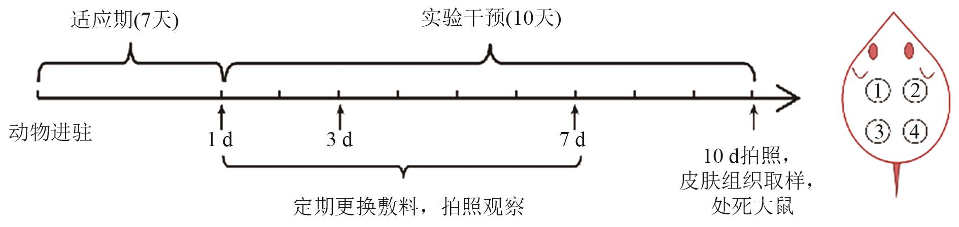
图1 大鼠全层创面愈合实验的实验流程图
Fig. 1 Flowchart of the rat full-thickness wound healing experiment
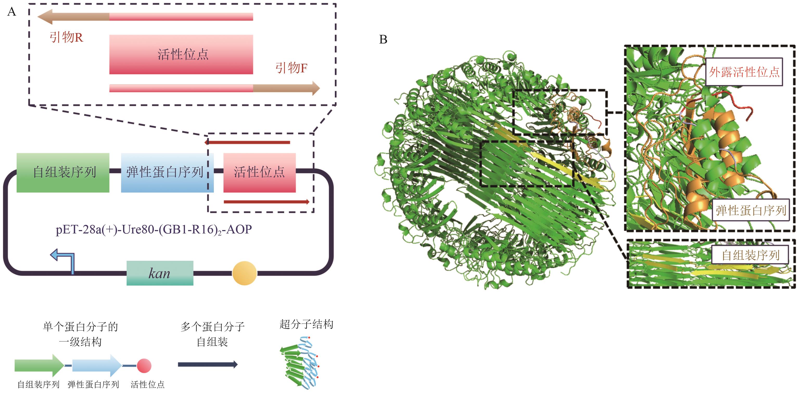
图2 基于自组装弹性蛋白序列构建抗氧化蛋白A:通过反向PCR构建重组蛋白表达质粒方法示意图。B:单个蛋白分子的一级结构以及自组装形成水凝胶结构的示意图。C:AlphaFold 3模拟自组装后蛋白的结构图,黄色部分为其中一个蛋白分子的自组装序列,橙色部分为弹性蛋白序列,红色部分为外露的活性位点
Fig. 2 Construction of antioxidant proteins based on self-assembling elastin-like sequencesA: Schematic diagram of recombinant protein expression plasmid construction via inverse PCR. B: Schematic diagram of the primary structure of a single protein molecule and the self-assembled hydrogel structure. C: Structure diagram of the self-assembled protein predicted by AlphaFold 3. The yellow region represents the self-assembling sequence of one protein molecule, the orange region represents the elastin-like sequence, and the red region represents the exposed active site
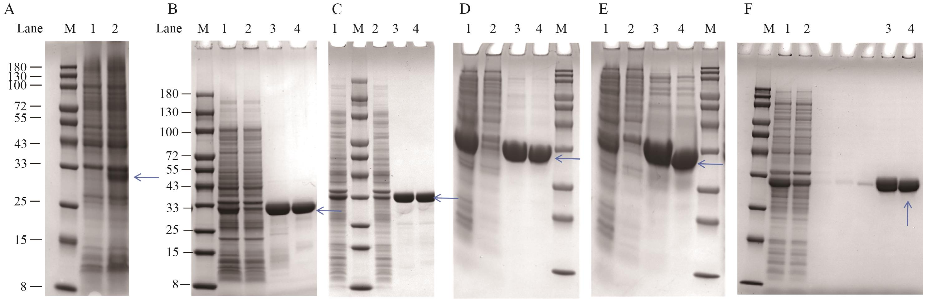
图3 蛋白表达与纯化的蛋白胶图A:蛋白发酵时,诱导大肠杆菌表达前后蛋白胶图。M:Marker;1:诱导前;2:诱导后。 B-E:分别为80-2-MPH、80-2-PP、80-2-RP1、80-2-Ⅳ5C、80-2-ABA蛋白纯化过程的蛋白胶图,分子量均为27 kD左右。M:Marker;1:细胞破碎液;2:流穿液;3:洗杂后镍柱洗脱蛋白;4:脱盐后蛋白
Fig. 3 Protein expression and purification gel imagesA: Gel images of protein expression in E. coli before and after induction during protein fermentation. M: Marker; 1: before induction; 2: after induction. B-E: Gel images showing the purification of 80-2-MPH, 80-2-PP, 80-2-RP1, 80-2-Ⅳ5C, and 80-2-ABA, respectively. The molecular weight of each protein was approximately 27 kD. M: Marker; 1: cell lysate; 2: flow-through; 3: Ni-column eluted protein after washing; 4: desalted protein
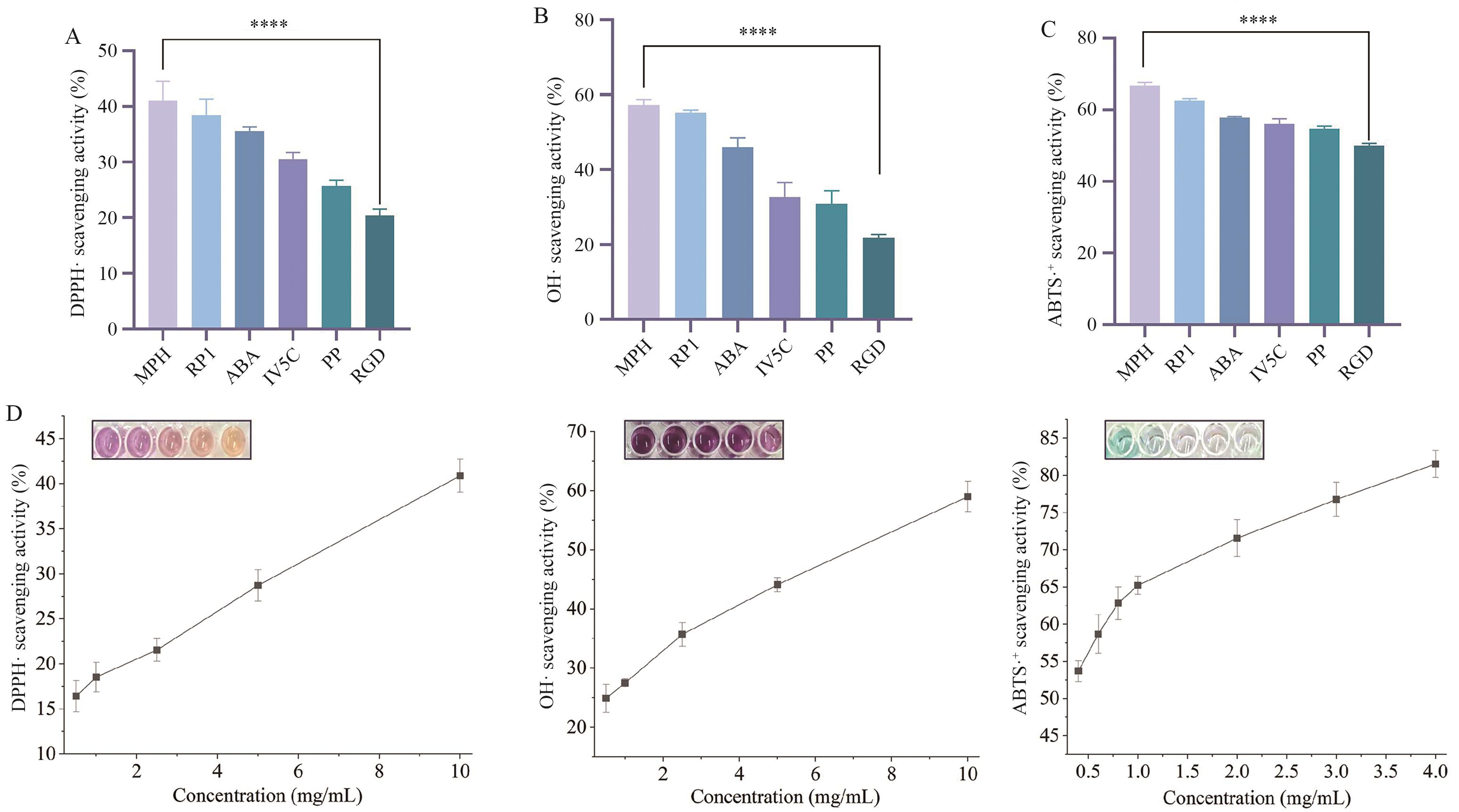
图4 化学实验表征不同蛋白的抗氧化性质A:不同蛋白DPPH自由基清除率。B:ABTS自由基清除率。C:不同蛋白·OH清除率。D:不同浓度80-2-MPH的自由基清除能力(照片为自由基清除效果照片)。****P<0.000 1,数据均表示为平均值±标准差(n=3),下同
Fig. 4 Chemical experiment characterizes the antioxidant properties of different proteinsA: DPPH· scavenging rate of different proteins. B: ABTS+· free radical scavenging rate. C: ·OH scavenging rate of different proteins. D: Free radical scavenging ability of different concentrations of 80-2-MPH. The photos show the effect of free radical scavenging. ****P<0.000 1, data are reported as mean ± SD (n=3), the same below
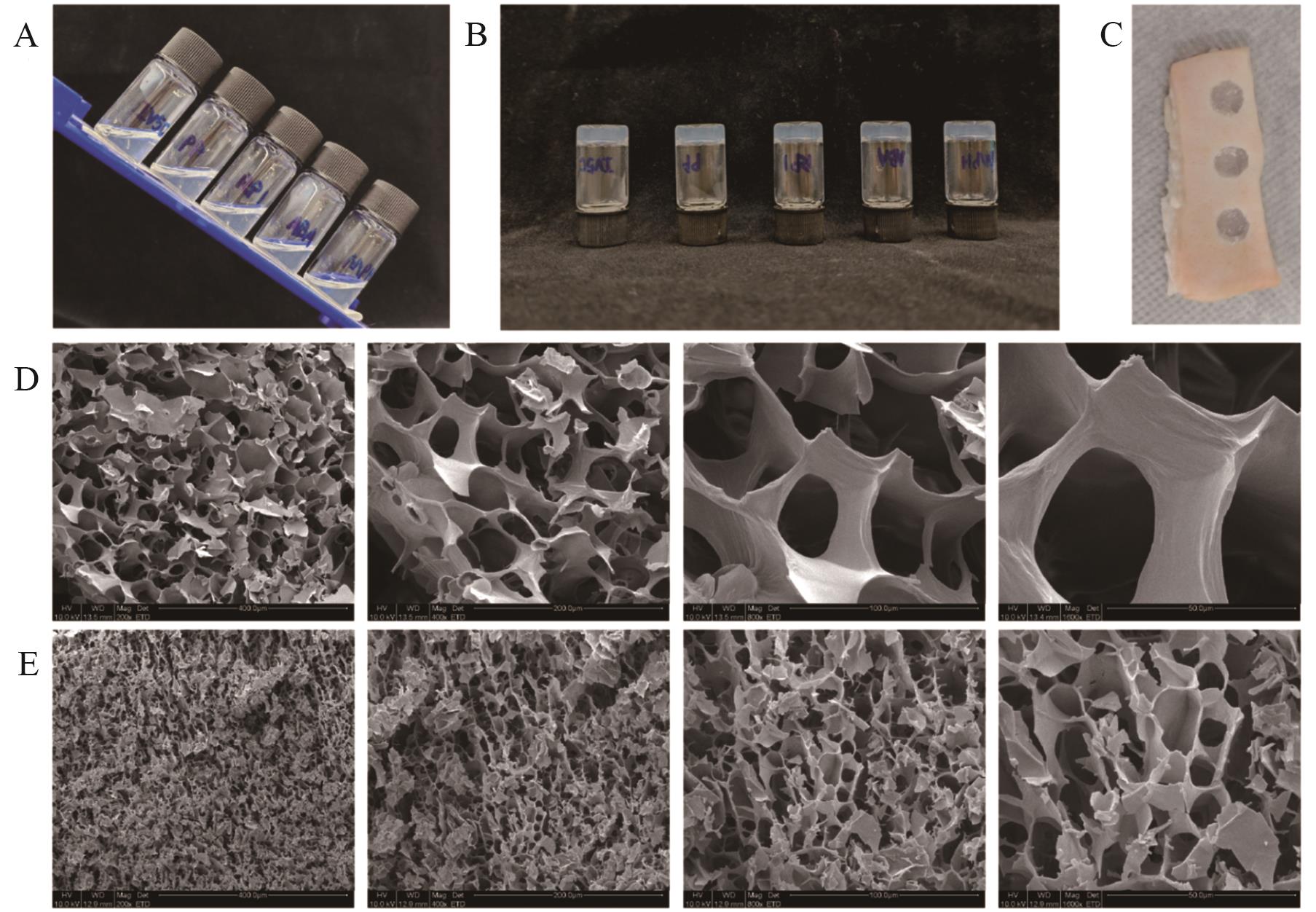
图5 水凝胶成胶性质的表征A:不同蛋白按1%(w/v)溶解于生理盐水,未成胶。从左到右分别为80-2-Ⅳ5C,80-2-PP,80-2-RP1,80-2-ABA和80-2-MPH。B:蛋白静置后自组装成水凝胶。C:浓度为4%(w/v)蛋白成胶后用注射器添加至猪皮缺损处,模拟蛋白作为伤口敷料应用时的场景。D:扫描电子显微镜观察5%(w/v)浓度水凝胶的网络结构。E:扫描电子显微镜观察10%(w/v)浓度水凝胶的网络结构
Fig. 5 Characterization of hydrogel gelation propertiesA: Different proteins were dissolved in physiological saline at a concentration of 1% (w/v), and no gelation occurred. From left to right: 80-2-Ⅳ5C, 80-2-PP, 80-2-RP1, 80-2-ABA, and 80-2-MPH. B: The protein self-assembled into a hydrogel after standing. C: After gelation at 4% (w/v), the protein hydrogel was applied to a porcine skin defect using a syringe to mimic its use as a wound dressing. D: Scanning electron microscopy images showing the network structure of the hydrogel at 5% (w/v). E: Scanning electron microscopy images showing the network structure of the hydrogel at 10% (w/v)
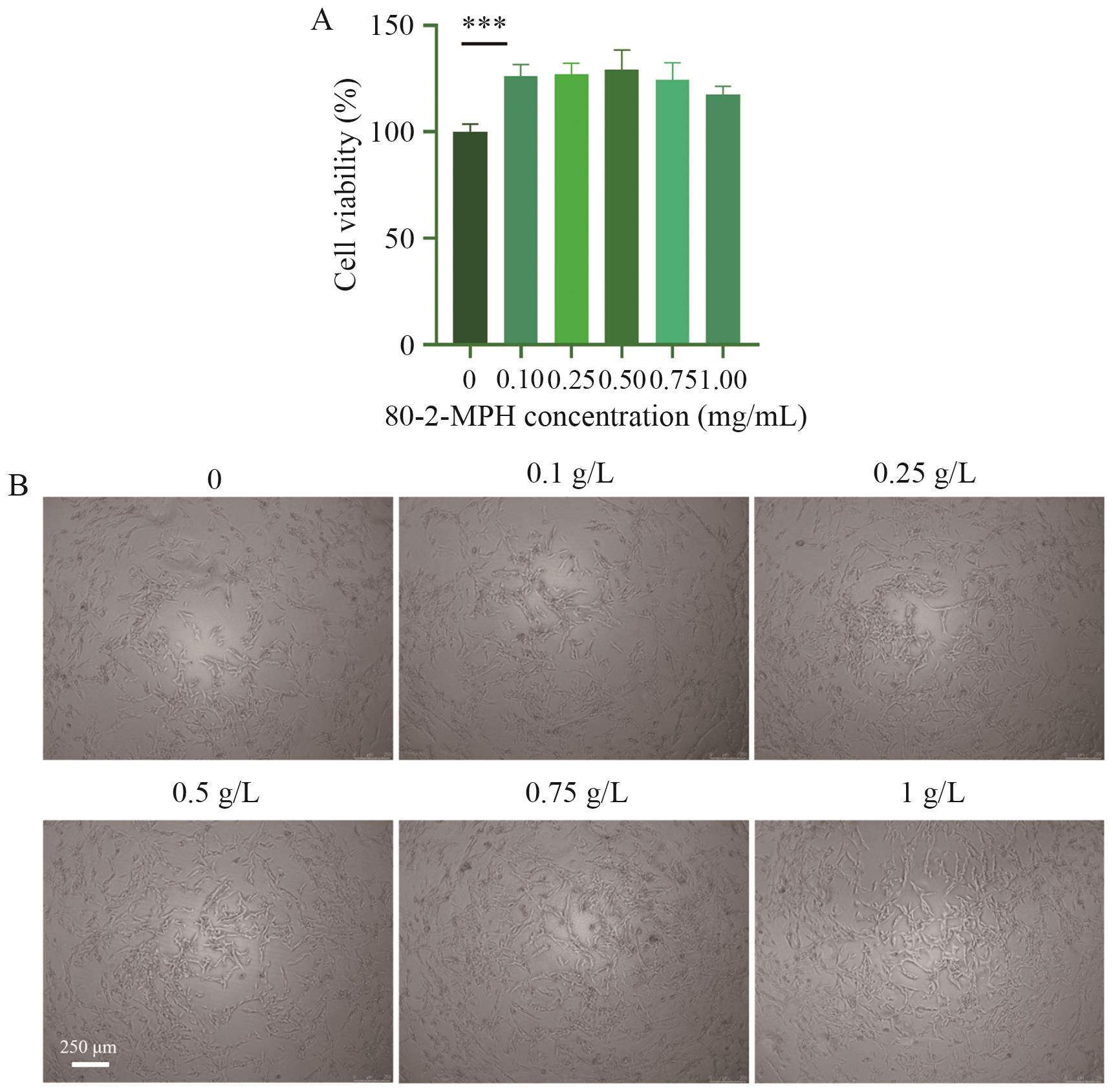
图6 80-2-MPH蛋白的生物活性A:80-2-MPH的生物相容性验证。含有不同浓度80-2-MPH的完全培养基培养细胞,CCK-8试剂盒检测细胞活力。B:显微镜观察各组细胞形态。***P<0.001
Fig. 6 Biological activity of 80-2-MPH proteinA: Verification of the biocompatibility of 80-2-MPH. Cells were cultured in complete medium containing different concentrations of 80-2-MPH, and cell viability was detected using the CCK-8 kit. B: Scanning electron microscopy images showing cell morphology in each group. ***P<0.001
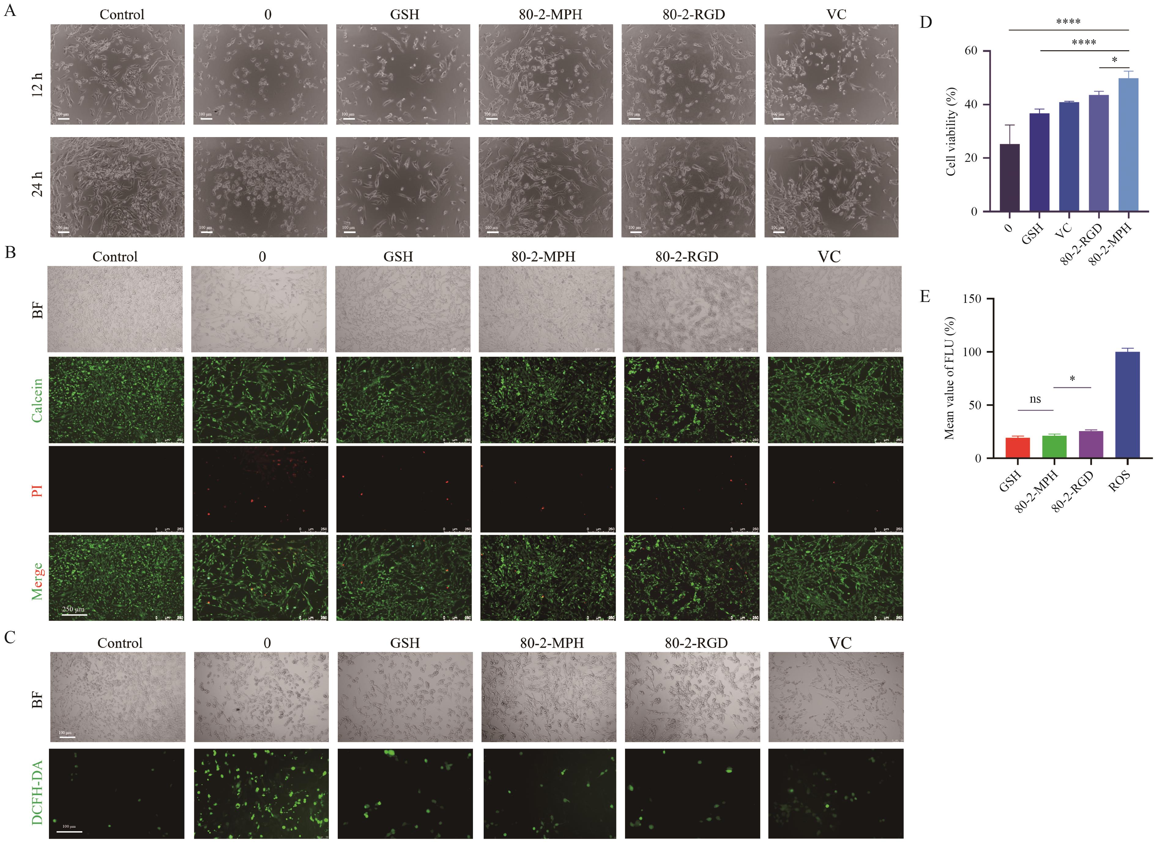
图7 80-2-MPH对细胞氧化损伤的修复作用以及对细胞中活性氧的清除作用A:损伤后修复12 h和24 h的细胞形态。 B:修复12 h时细胞活死染。C:DCFH-DA检测细胞中的活性氧。D:修复12 h时CCK-8测定各组的细胞活性。数据均表示为平均值±标准差(n=4)。E:流式细胞仪检测细胞中的相对活性氧含量。*P<0.05
Fig. 7 Effect of 80-2-MPH on the repair of cellular oxidative damage and the scavenging of intracellular reactive oxygen speciesA: Cell morphology at 12 h and 24 h after damage repair. B: Live/dead cell staining at 12 h after damage repair. C: Detection of reactive oxygen species in cells using DCFH-DA. D: Cell viability was measured by CCK-8 assay in each group at 12 h of repair. Data are reported as means ± SD (n=4). E: Detection of relative reactive oxygen species content in cells using flow cytometry. *P<0.05
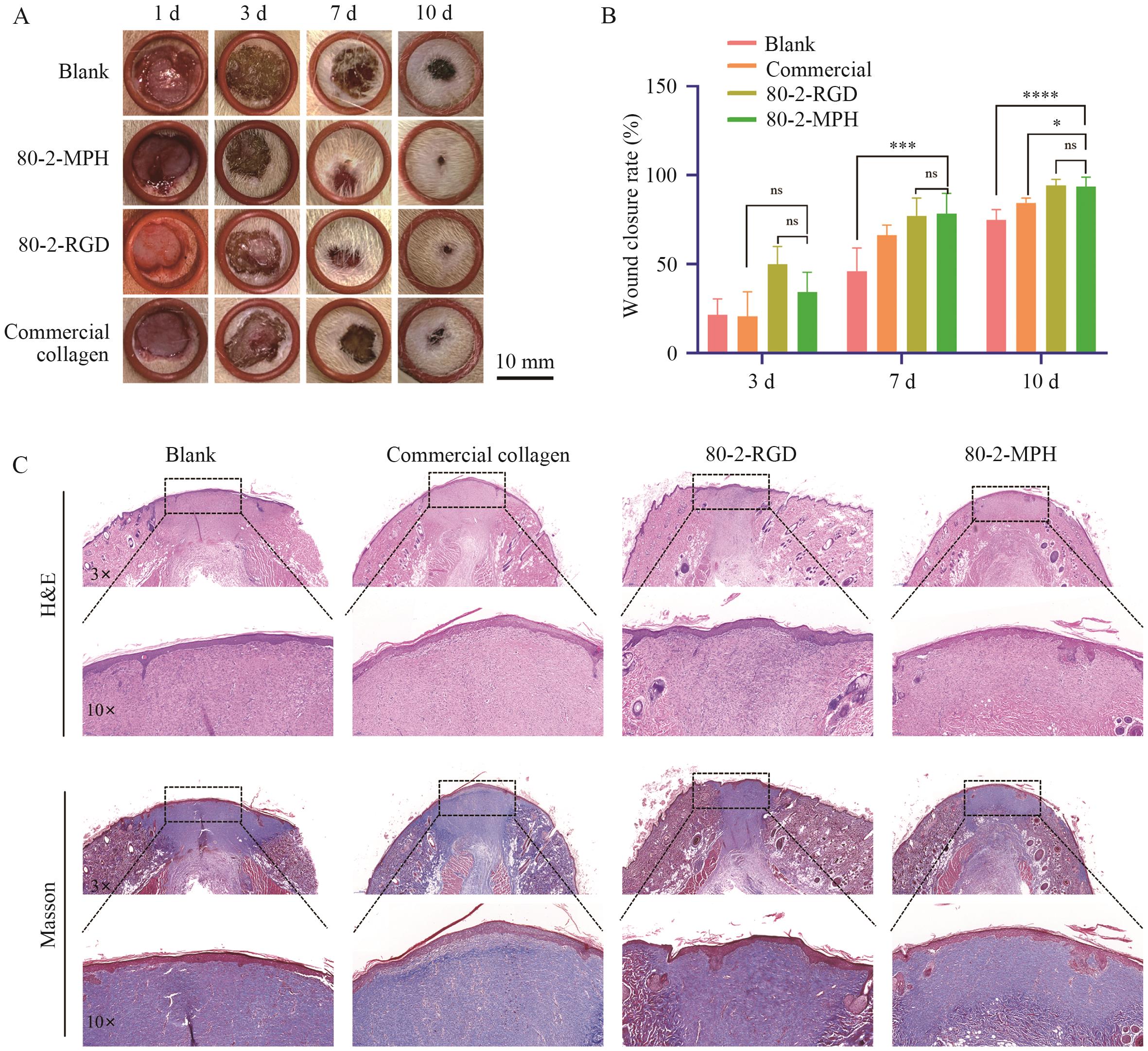
图8 大鼠全层创面闭合情况A:定时记录不同组大鼠的伤口愈合情况。B:不同组大鼠伤口闭合率计算。数据均表示为平均值±标准差(n=6)。C:皮肤样品的H&E染色和Masson染色
Fig. 8 Closure of full-thickness wounds in ratsA: Wound healing in different groups of rats was recorded at regular intervals. B: Wound closure rate in different groups of rats. Data are reported as means ± SD (n=6). C: H&E staining and Masson staining of skin samples
| [1] | Sun J, Su JJ, Ma C, et al. Fabrication and mechanical properties of engineered protein-based adhesives and fibers [J]. Adv Mater, 2020, 32(6): 1906360. |
| [2] | Zhao ZL, Deng JJ, Fan DD. Green biomanufacturing in recombinant collagen biosynthesis: trends and selection in various expression systems [J]. Biomater Sci, 2023, 11(16): 5439-5461. |
| [3] | Xu R, Luo GX, Xia HS, et al. Novel bilayer wound dressing composed of silicone rubber with particular micropores enhanced wound re-epithelialization and contraction [J]. Biomaterials, 2015, 40: 1-11. |
| [4] | Ahmed SR, Mostafa EM, Musa A, et al. Wound healing and antioxidant properties of Launaea procumbens supported by metabolomic profiling and molecular docking [J]. Antioxidants, 2022, 11(11): 2258. |
| [5] | Mouthuy PA, Snelling SJB, Dakin SG, et al. Biocompatibility of implantable materials: an oxidative stress viewpoint [J]. Biomaterials, 2016, 109: 55-68. |
| [6] | Dan A, Sharma D, Singh H, et al. A dynamically crosslinked, self-adapting, injectable gelatin-chondroitin sulfate hydrogel with antibacterial and antioxidant properties for treatment of deep and irregular wounds [J]. J Mater Chem B, 2025, 13(25): 7293-7310. |
| [7] | Ding RJ, Xu XC, Ding HY, et al. A multifunctional injectable hydrogel accelerates hemostasis and wound healing via synergistic tissue adhesion, antimicrobial activity, and ROS scavenging [J]. ACS Appl Mater Interfaces, 2025, 17(44): 60255-60268. |
| [8] | Li Y, Sun ZH, Chai SR, et al. Tissue-like soft dual-network hydrogel patch with instant/tunable adhesion and microenvironment modulation for infected wound healing [J]. ACS Nano, 2025, 19(30): 27714-27730. |
| [9] | Kumar AG, Nair A, Sreejith S, et al. Biocompatible PVA-chitosan hydrogel enriched with collagen hydrolysate and Couroupita guianensis flower extract for enhanced wound-healing and antibacterial activity [J]. Polym Bull, 2025, 82(13): 7747-7769. |
| [10] | Yang MY, Liu PY, Cheng P, et al. Naturally derived hydrogel with antioxidant, angiogenesis and photothermal effect to accelerate infected diabetic wound healing and reduce scar formation [J]. Theranostics, 2025, 15(18): 9838-9861. |
| [11] | Stachowiak M, Mlynarczyk DT, Dlugaszewska J. Wondrous yellow molecule: are hydrogels a successful strategy to overcome the limitations of curcumin? [J]. Molecules, 2024, 29(8): 1757. |
| [12] | Guo QY, Zhang MM, Zheng JY, et al. A multifunctional MXene@CeO2-enhanced hydrogel dressing for synergistic photothermal antibacterial and antioxidative therapy in wound healing [J]. Adv Healthc Mater, 2025, 14(23): 2500656. |
| [13] | Wang WL, Lai YH, Huang CH, et al. Lumbrokinase-containing gelatin nanofibers with multiple bioactivities for effective skin wound healing [J]. Mater Today Bio, 2025, 32: 101713. |
| [14] | Yang M, Chen X, Chen ZX, et al. Thermoresponsive antioxidant metal-free carbon nanodot hydrogel: an effective therapeutic approach for ocular surface disease [J]. Sci Adv, 2025, 11(30): eadt8775. |
| [15] | Álvarez Z, Kolberg-Edelbrock AN, Sasselli IR, et al. Bioactive scaffolds with enhanced supramolecular motion promote recovery from spinal cord injury [J]. Science, 2021, 374(6569): 848-856. |
| [16] | Ogrodnik M. Aging: the wound that never starts healing [J]. Nat Commun, 2025, 16: 8732. |
| [17] | Yoshitomi T, Hirayama A, Nagasaki Y. The ROS scavenging and renal protective effects of pH-responsive nitroxide radical-containing nanoparticles [J]. Biomaterials, 2011, 32(31): 8021-8028. |
| [18] | Wang LL, Xue B, Zhang X, et al. Extracellular matrix-mimetic intrinsic versatile coating derived from marine adhesive protein promotes diabetic wound healing through regulating the microenvironment [J]. ACS Nano, 2024, 18(22): 14726-14741. |
| [19] | Wu JJ, Zhou L, Peng H, et al. A general and convenient peptide self-assembling mechanism for developing supramolecular versatile nanomaterials based on the biosynthetic hybrid amyloid-resilin protein [J]. Adv Mater, 2024, 36(4): 2304364. |
| [20] | 王兆石, 王召君, 董明盛, 等. 超分子弹性蛋白的促细胞增殖及抗氧化能力研究 [J]. 食品与生物技术学报, 2025, 44(1): 132-142. |
| Wang ZS, Wang ZJ, Dong MS, et al. Cell proliferation-promoting effect and antioxidant capacity of supramolecular elastin [J]. J Food Sci Biotechnol, 2025, 44(1): 132-142. | |
| [21] | Xu XQ, Lai R. The chemistry and biological activities of peptides from amphibian skin secretions [J]. Chem Rev, 2015, 115(4): 1760-1846. |
| [22] | Chi CF, Wang B, Deng YY, et al. Isolation and characterization of three antioxidant pentapeptides from protein hydrolysate of monkfish (Lophius litulon) muscle [J]. Food Res Int, 2014, 55: 222-228. |
| [23] | Harnedy PA, O’Keeffe MB, FitzGerald RJ. Fractionation and identification of antioxidant peptides from an enzymatically hydrolysed Palmaria palmata protein isolate [J]. Food Res Int, 2017, 100: 416-422. |
| [24] | Zhang M, Mu TH, Sun MJ. Purification and identification of antioxidant peptides from sweet potato protein hydrolysates by Alcalase [J]. J Funct Foods, 2014, 7: 191-200. |
| [25] | Wang QQ, Wang LY, Huang ZL, et al. Abalone peptide increases stress resilience and cost-free longevity via SKN-1-governed transcriptional metabolic reprogramming in C. elegans [J]. Aging Cell, 2024, 23(2): e14046. |
| [26] | 刘文睿. 超分子弹性蛋白水凝胶的合成生物设计与功能研究 [D]. 无锡: 江南大学, 2025. |
| Liu WR. Synthesis, biological design and functional study of supramolecular elastin hydrogel [D]. Wuxi: Jiangnan University, 2025. | |
| [27] | Züger S, Iwai H. Intein-based biosynthetic incorporation of unlabeled protein tags into isotopically labeled proteins for NMR studies [J]. Nat Biotechnol, 2005, 23(6): 736-740. |
| [28] | Xiao F, Xu T, Lu BY, et al. Guidelines for antioxidant assays for food components [J]. Food Front, 2020, 1(1): 60-69. |
| [29] | Zhao MY, Xiang JW, Meng Y, et al. Astragalus polysaccharide hydrogels with drug-carrying super self-assembly from natural herbs promote wound healing [J]. ACS Nano, 2025, 19(23): 21571-21588. |
| [30] | Wang CH, Yu DY, Liu W, et al. Bioinspired co-assembled hydrogels constructed from marine self-assembling peptides and polyphenol network: antioxidant and infected wound healing [J]. Antioxidants, 2025, 14(7): 785. |
| [31] | Yang FM, Wang N, Cui SH, et al. Marine-inspired self-assembled peptide hydrogel spatiotemporal treatment of bacterial-infected wounds [J]. Chem Eng J, 2025, 519: 165573. |
| [32] | Yang D, Zhao W, Zhang SY, et al. Dual self-assembly of puerarin and silk fibroin into supramolecular nanofibrillar hydrogel for infected wound treatment [J]. Adv Healthc Mater, 2024, 13(19): 2400071. |
| [33] | Sunil A, Hernández Rico YG, Abioye RO, et al. Biofunctional peptide hydrogel: self-assembly, rheological properties, and in vivo antioxidant activity of ovalbumin-derived peptide (VLVNAIVFKGL) [J]. J Agric Food Chem, 2025, 73(10): 5961-5969. |
| [34] | Chen JY, Wang X, Yuan XM, et al. Bioinspired multifunctional hydrogel loaded with netrin-1 for diabetic wound healing [J]. ACS Appl Mater Interfaces, 2025, 17(39): 54525-54536. |
| [35] | Fang Y, Xu K, Feng XC, et al. Bioactive self-adaptive hydrogel platform for integrated electrical stimulation therapy and clinical monitoring of postoperative wound [J]. ACS Sens, 2025, 10(10): 7757-7777. |
| [36] | Forman HJ, Zhang HQ. Targeting oxidative stress in disease: promise and limitations of antioxidant therapy [J]. Nat Rev Drug Discov, 2021, 20(9): 689-709. |
| [37] | Ramachandran V, Mohanasundaram T, Tiwari R, et al. Nrf2 mediated heme oxygenase-1 activation contributes to Diabetic Wound healing-an overview [J]. Drug Res (Stuttg), 2022, 72(9): 487-495. |
| [38] | Huang W, Guo QR, Wu HR, et al. Engineered exosomes loaded in intrinsic immunomodulatory hydrogels with promoting angiogenesis for programmed therapy of diabetic wounds [J]. ACS Nano, 2025, 19(14): 14467-14483. |
| [39] | Zhang W, Zhai WX, Xu M, et al. Constructing reinforced flexible wood-based hydrogels leveraging the ordered structure of wood for potential wound treatment [J]. ACS Sustainable Chem Eng, 2025, 13(15): 5689-5702. |
| [40] | Xue HQ, Gu QC, Zhong XQ, et al. Polysaccharide micelle hydrogel delivery Ginkgolide B for wound healing [J]. Mater Des, 2025, 257: 114505. |
| [41] | Wang ZX, Liu J, Chen QY, et al. Bioactive glycyrrhizic acid ionic liquid self-assembled nanomicelles for enhanced transdermal delivery of anti-photoaging signal peptides [J]. Adv Sci, 2025, 12(8): 2412581. |
| [42] | Xue B. Efficacy and cellular mechanism of biomimetic marine adhesive protein-based coating against skin photoaging [J]. Adv Healthc Mater, 2026, 15(10): 2402019. |
| [43] | Lian CX, Liu JW, Wei WY, et al. Mg-gallate metal-organic framework-based sprayable hydrogel for continuously regulating oxidative stress microenvironment and promoting neurovascular network reconstruction in diabetic wounds [J]. Bioact Mater, 2024, 38: 181-194. |
| [1] | 任文斌, 陈登科, 吴翠翠. 花生转录因子AhZHD9的生物信息学分析及耐盐性研究[J]. 生物技术通报, 2026, 42(9): 297-305. |
| [2] | 刘君良, 袁晓霞, 乔美玲, 杨冰洁, 于秀敏, 穆莎茉莉, 冀照君. 基因aotM与trmD调控根瘤菌盐碱耐受性、抗氧化酶活性及细胞膜氧化损伤[J]. 生物技术通报, 2026, 42(8): 323-330. |
| [3] | 董亚茹, 修妤, 赵东晓, 朱红, 朱琳, 刘惠芬. 基于转录组的桑树GRAS基因家族鉴定及MnGRAS22盐胁迫响应分析[J]. 生物技术通报, 2026, 42(7): 193-203. |
| [4] | 李博宇, 蹇春晨, 侯佳俊, 钱明岩, 杨文钦, 章圣龙, 藏传刚, 马福开, 郑龙, 关宏. 复合菌剂QY12-A4对玉米秸秆的降解及其产物中抗氧化木质素的分离与纯化[J]. 生物技术通报, 2026, 42(7): 292-303. |
| [5] | 李灿妮, 潘炜松, 吴泰茹, 吴川, 曾宝强, 李伟展. 胚乳生物反应器表达重组蛋白的研究进展[J]. 生物技术通报, 2026, 42(7): 67-80. |
| [6] | 王冰冰, 朱迪, 杨生龙, 李艺林, 杨琪, 贺苗苗. 外源褪黑素对晚疫病胁迫下马铃薯抗病性的影响[J]. 生物技术通报, 2026, 42(6): 107-115. |
| [7] | 莫言玲, 刘源橼, 裴佳玲, 樊襄曼, 邓兴连, 曾静, 刘义华. 外源硒预处理缓解茎瘤芥幼苗镉毒害的机制研究[J]. 生物技术通报, 2026, 42(6): 304-313. |
| [8] | 吴夏明, 刘传和, 贺涵, 周陈平, 杨敏, 邝瑞彬, 徐泽, 魏岳荣. 草莓耐高温分子响应机制的研究进展[J]. 生物技术通报, 2026, 42(6): 175-185. |
| [9] | 任泓宇, 庞翠萍, 古阳, 周佳海. 多策略协同提升蛋白质晶体的衍射分辨率[J]. 生物技术通报, 2026, 42(1): 76-85. |
| [10] | 陈强, 于璎霏, 张颖, 张冲. 茉莉酸甲酯对薄皮甜瓜‘绿宝石’采后冷害的调控[J]. 生物技术通报, 2025, 41(9): 105-114. |
| [11] | 蔡如凤, 杨宇轩, 于基正, 李佳楠. 人工智能重塑蛋白质工程:从结构解析到合成生物学的算法革命[J]. 生物技术通报, 2025, 41(8): 1-10. |
| [12] | 程珊, 王会伟, 陈晨, 朱雅婧, 李春鑫, 别海, 王树峰, 陈献功, 张向歌. 油莎豆MYB转录因子基因CeMYB154克隆及耐盐功能分析[J]. 生物技术通报, 2025, 41(6): 218-228. |
| [13] | 佟德利, 张馨, 陈佳庆, 贺海升. 蓝莓内生细菌的分离及其对上海青铝胁迫的缓解作用[J]. 生物技术通报, 2025, 41(4): 302-311. |
| [14] | 鲁天怡, 李爱朋, 费强. 生物合成聚乳酸研究进展[J]. 生物技术通报, 2025, 41(4): 47-60. |
| [15] | 钱祺, 王增辉, 孙荣华, 罗英智, 苏良辰. 花生蛋白磷酸酶AhPDCP37的抗旱性功能研究[J]. 生物技术通报, 2025, 41(3): 98-103. |
| 阅读次数 | ||||||
|
全文 |
|
|||||
|
摘要 |
|
|||||
